-
Články
Reklama
- Vzdělávání
- Časopisy
Top články
Nové číslo
- Témata
Reklama- Videa
- Podcasty
Nové podcasty
Reklama- Kariéra
Doporučené pozice
Reklama- Praxe
ReklamaAllelic Variation and Differential Expression of the mSIN3A Histone Deacetylase Complex Gene Promote Mammary Tumor Growth and Metastasis
Accumulating evidence suggests that breast cancer metastatic progression is modified by germline polymorphism, although specific modifier genes have remained largely undefined. In the current study, we employ the MMTV-PyMT transgenic mouse model and the AKXD panel of recombinant inbred mice to identify AT–rich interactive domain 4B (Arid4b; NM_194262) as a breast cancer progression modifier gene. Ectopic expression of Arid4b promoted primary tumor growth in vivo as well as increased migration and invasion in vitro, and the phenotype was associated with polymorphisms identified between the AKR/J and DBA/2J alleles as predicted by our genetic analyses. Stable shRNA–mediated knockdown of Arid4b caused a significant reduction in pulmonary metastases, validating a role for Arid4b as a metastasis modifier gene. ARID4B physically interacts with the breast cancer metastasis suppressor BRMS1, and we detected differential binding of the Arid4b alleles to histone deacetylase complex members mSIN3A and mSDS3, suggesting that the mechanism of Arid4b action likely involves interactions with chromatin modifying complexes. Downregulation of the conserved Tpx2 gene network, which is comprised of many factors regulating cell cycle and mitotic spindle biology, was observed concomitant with loss of metastatic efficiency in Arid4b knockdown cells. Consistent with our genetic analysis and in vivo experiments in our mouse model system, ARID4B expression was also an independent predictor of distant metastasis-free survival in breast cancer patients with ER+ tumors. These studies support a causative role of ARID4B in metastatic progression of breast cancer.
Published in the journal: . PLoS Genet 8(5): e32767. doi:10.1371/journal.pgen.1002735
Category: Research Article
doi: https://doi.org/10.1371/journal.pgen.1002735Summary
Accumulating evidence suggests that breast cancer metastatic progression is modified by germline polymorphism, although specific modifier genes have remained largely undefined. In the current study, we employ the MMTV-PyMT transgenic mouse model and the AKXD panel of recombinant inbred mice to identify AT–rich interactive domain 4B (Arid4b; NM_194262) as a breast cancer progression modifier gene. Ectopic expression of Arid4b promoted primary tumor growth in vivo as well as increased migration and invasion in vitro, and the phenotype was associated with polymorphisms identified between the AKR/J and DBA/2J alleles as predicted by our genetic analyses. Stable shRNA–mediated knockdown of Arid4b caused a significant reduction in pulmonary metastases, validating a role for Arid4b as a metastasis modifier gene. ARID4B physically interacts with the breast cancer metastasis suppressor BRMS1, and we detected differential binding of the Arid4b alleles to histone deacetylase complex members mSIN3A and mSDS3, suggesting that the mechanism of Arid4b action likely involves interactions with chromatin modifying complexes. Downregulation of the conserved Tpx2 gene network, which is comprised of many factors regulating cell cycle and mitotic spindle biology, was observed concomitant with loss of metastatic efficiency in Arid4b knockdown cells. Consistent with our genetic analysis and in vivo experiments in our mouse model system, ARID4B expression was also an independent predictor of distant metastasis-free survival in breast cancer patients with ER+ tumors. These studies support a causative role of ARID4B in metastatic progression of breast cancer.
Introduction
Breast cancer remains the most commonly diagnosed malignancy among women in the United States [1]. Because the vast majority of breast cancer related mortality is attributable to disseminated metastatic disease, a clear need exists to identify factors that modulate breast cancer metastatic progression. In addition to acquired somatic mutations, there is accumulating evidence that the genetic background on which a tumor arises can influence disease progression [2]. Identifying and characterizing metastasis susceptibility genes would provide additional insights into the mechanisms associated with tumor dissemination and growth, leading not only to better understanding of this complex process but also ultimately to new targets and strategies for clinical intervention.
Due to the complex interactions between inherited factors and somatic mutations in metastatic progression, as well as the genetic complexity of human populations, identification of inherited susceptibility genes directly in human populations is difficult. To circumvent this our laboratory has chosen to apply a systems genetics approach on a mouse model of metastatic luminal breast cancer, the FVB/N-TgN(MMTV-PyMT)634Mul (MMTV-PyMT) transgenic model. The MMTV-PyMT transgenic mouse model, which expresses the polyoma virus middle T antigen under the control of the mouse mammary tumor virus promoter, rapidly develops tumors in approximately 100% of female mammary glands and >85% of these animals develop pulmonary metastases by 14 weeks of age. When the MMTV-PyMT model is bred onto a variety of different mouse strains, the F1 progeny display broad and strain-dependent heterogeneity in primary tumor latency, primary tumor growth rate and lung metastatic density [2].
Two strains, the highly metastatic AKR/J and poorly metastatic DBA/2J, were found to have a 20-fold difference in their metastatic capacity but no significant difference in any other measured tumor phenotype. These strains were also the progenitor strains for the AKXD recombinant inbred panel of mice, which consists of more than 20 substrains that are composites of the original parental strains AKR/J and DBA/2J. The MMTV-PyMT model was therefore bred to 18 different AKXD strains, the F1 mice were phenotyped with respect to primary tumor latency and burden and lung metastatic density, and the phenotypes were compared to haplotype maps of the AKXD strains to determine quantitative trait loci (QTLs) associated with mammary tumor progression [3]. Subsequently, RNA was also harvested from F1 tumors and gene expression analysis was performed to define individual genes whose expression correlated with progression [4].
In this study we have utilized these resources to identify Arid4b as a novel candidate metastasis susceptibility gene. Although the precise molecular functions of ARID4B are unknown, it has been shown to associate with the SIN3A histone deacetylase (HDAC) complex [5]. As predicted by the genetic linkage and gene expression data, higher expression of Arid4b is associated with more rapid tumor growth in animal models, as well as increased tumor cell motility and invasion. These effects are associated with differential binding of the AKR and DBA alleles of ARID4B to HDAC complex members mSIN3A and mSDS3. ARID4B was also found to bind the mSIN3A-associated breast cancer metastasis suppressor protein BRMS1. Stable shRNA-mediated knockdown of Arid4b significantly inhibited the pulmonary metastatic efficiency of orthotopic mammary tumors without inhibiting primary tumor growth. Consistent with impaired metastasis in the Arid4b knockdown lines was decreased expression of a recently described metastasis-predictive gene network [6]. High expression of ARID4B was associated with an approximately 2-fold increased risk of metastatic progression in human breast cancer patients who were lymph node negative at diagnosis. Taken together these results demonstrate a causal role for Arid4b in tumor growth and metastatic progression and suggest that mechanisms of action involve modification of epigenetic state via the mSIN3A complex and regulation of the conserved Tpx2 gene network.
Results
Identification of Arid4b as a potential tumor progression gene
Previously a cross between the highly metastatic PyMT model and the AKXD recombinant inbred (RI) panel was performed to map QTLs associated with inherited predisposition to developing pulmonary metastasis [3]. In addition to metastasis susceptibility loci on chromosomes 6 and 19, linkage analysis revealed a potential peak on proximal chromosome 13 (Figure S1). In a subsequent study, gene expression analysis was also carried out on these samples to examine the effect of varying metastatic genotypes on tumor transcriptional patterns [7]. To discover potential candidate genes that may affect metastatic predisposition, correlation analysis was performed using GeneNetwork [8] to identify genes whose differential expression was highly associated with metastasis. Upon integrating the data from these two studies we found that of the top ten genes most significantly associated with metastasis in our expression correlation analysis, two also mapped to potential QTLs: Ttc9c and Arid4b. The potential role of Ttc9c was investigated and no significant differences were detected with respect to orthotopic tumor growth or metastasis of 6DT1 mouse mammary carcinoma cells stably expressing Ttc9c compared to vector control cells (data not shown). Similarly, we detected no significant effects on tumor growth or metastasis when Ttc9c BAC transgenic mice were bred to the MMTV-PyMT model (data not shown). The most likely explanation for why Ttc9c did not pass our validation experiments is that its initial identification in our screens was a false positive owing to its close physical proximity on chromosome 19 to the metastasis modifier gene Sipa1 [9]. Our current studies have therefore focused on Arid4b, which maps within the chromosome 13 locus and whose mRNA expression was positively associated with metastatic disease and tumor growth (Figure 1), suggesting a possible causative role as a progression modifier.
Fig. 1. Arid4b expression is correlated with tumor growth and metastasis. 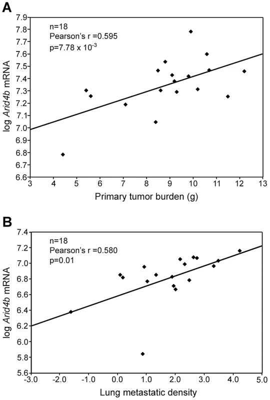
Gene expression microarray data from 18 of the AKXD recombinant inbred strains shows expression of Arid4b is positively correlated with tumor burden (A) and metastatic density (B). The formula used to calculate metastatic density was ln[(metastases/um2)×108]. Arid4b is both differentially expressed and polymorphic between AKR and DBA
To validate the potential differences in Arid4b expression between strains, microarray data from AKR and DBA normal tissues were examined [4]. Consistent with the AKXD RI results, Arid4b expression was 2.3-fold higher in thymus (p = 9.32×10⁻⁵, FDR = 0.0004) and 2.5-fold higher in bone marrow (p = 1.28×10⁻⁵, FDR = 0.0005) of DBA mice compared to AKR, suggesting that constitutional polymorphisms can influence Arid4b expression levels in normal tissues. Sequence analysis was also performed to both validate SNPs in the public database as well as identify potential new variants between the AKR and DBA alleles of Arid4b. Complete exon sequencing revealed that the DBA allele matched the consensus C57BL/6 sequence. Analysis of the AKR allele revealed numerous silent SNPs as well as polymorphisms encoding eleven amino acid substitutions, as shown in Figure 2. Interestingly, eight of these eleven polymorphisms are located in exon 22 and their encoded substitutions are densely clustered towards the C-terminal end between amino acids 1171 and 1198. These results are consistent with the possibility that inherited variation of Arid4b may contribute to tumor progression.
Fig. 2. Arid4b germline amino acid sequence variants. 
Substitutions in the AKR allele are shown relative to a consensus sequence that is identical between DBA, FVB, and C57Bl/6. The+symbol indicates conserved substitutions. Arid4b expression promotes primary tumor growth
Analysis of the data revealed that increased Arid4b expression and increased metastatic susceptibility were associated with the DBA rather than the AKR genotype at the chromosome 13 QTL. This result suggests that the DBA allele at this locus promoted metastatic progression relative to the AKR allele, and a series of in vitro and in vivo assays were performed to test this hypothesis. V5-tagged AKR and DBA alleles of Arid4b were ectopically expressed in the mouse mammary carcinoma cell line Met-1, which was originally derived from tumors arising in the MMTV-PyMT transgenic model [10]. Because the Met-1 line was derived from an FVB strain background, we also sequenced the FVB allele of Arid4b and found it to be identical to the DBA and C57BL/6 alleles. Cell lines were then identified that expressed the epitope tagged constructs at levels that were only two to three-fold higher than endogenous levels as measured by QRT-PCR (Figure S2A), consistent with the approximately two-fold range of Arid4b mRNA between high and low metastatic AKXD strains. Furthermore, the ectopically expressed AKR and DBA alleles were detected at approximately equal levels in our stable lines as assessed by western blots (Figure S2B).
Orthotopic implantation assays were then performed to examine the role of Arid4b expression in vivo (Figure 3A). By four weeks post-implantation, cells expressing the DBA allele formed tumors with a 2.6-fold larger mass compared to control cells (741 mg versus 284 mg; p = 6.08×10∧−7). The AKR allele expressing cells formed tumors with a median mass of 480 mg, which was significantly larger than control tumors (p = 0.010) but significantly smaller than the DBA cohort (p = 7.73×10∧−3), consistent with our previous genetic analysis and our in vitro studies.
Fig. 3. Ectopic expression of Arid4b increases orthotopic tumor growth, tumor cell migration, and invasion. 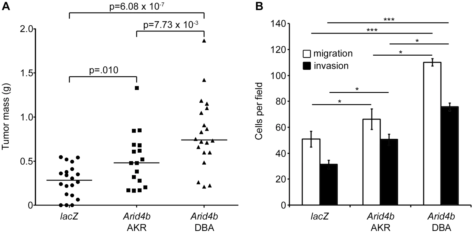
Scatter plot bars represent median tumor mass (A) four weeks after orthotopic implantation of 10∧5 cells stably expressing lacZ (n = 20), Arid4b AKR allele (n = 17) or Arid4b DBA allele (n = 20). Migrating and invading cells (B) were counted in five 400× fields per experiment and data are presented as the mean ± standard error of three independent experiments. Statistical significance was determined using Kruskal-Wallis tests followed by Conover-Inman post hoc tests. *, p<0.05; ***, p<0.001. DBA Arid4b promotes increased in vitro tumor invasion and migration compared to AKR
In vitro assays were performed to address the potential affect of the Arid4b polymorphisms on tumor cell behavior. In vitro growth assays demonstrated no significant difference in proliferation between cells expressing the DBA or AKR alleles or control cells (data not shown). In contrast, ectopic expression of either allele significantly increased the abilities of Met-1 cells to migrate through a porous membrane and to invade through Matrigel, compared to control cells expressing lacZ (Figure 3B). Notably, Met-1 cells stably expressing the DBA allele were significantly more migratory and invasive than those expressing the AKR allele. Since both cell lines express the epitope-tagged construct at approximately the same level, these results suggest a potential functional consequence for the amino acid substitutions present between the two variants in addition to the effects associated with differential expression.
Knockdown of Arid4b inhibits pulmonary metastasis
Because Met-1 cells are poorly metastatic in our laboratory, and because we were unable to stably overexpress Arid4b in several more aggressive mouse breast cancer cell lines, we adopted a knockdown strategy to examine the role of Arid4b in lung metastasis in vivo. To this end, the highly metastatic 6DT1 cell line [11] was transduced with five lentiviral shRNAs targeting Arid4b, or a scrambled control, and knockdown of ARID4B protein was evaluated using western blots (Figure 4A) and densitometry (Figure 4B) to select stable shRNA lines for in vivo studies. No significant knockdown was observed using the scrambled control shRNA. Cells stably transduced with Arid4b shRNAs designated H3 and H4 expressed 81% and 85% less ARID4B protein, respectively, compared to controls, and were therefore selected for further in vivo study.
Fig. 4. Stable shRNA–mediated knockdown of Arid4b inhibits lung metastasis. 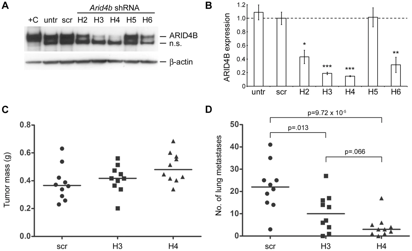
ARID4B protein levels were analyzed by western blot to determine level of knockdown in cell lines stably expressing different Arid4b shRNAs. Experiment was performed in triplicate and a representative blot is shown in panel A. Lysate from 293 cells transiently transfected with Arid4b was used as a positive control (+C). Untransduced 6DT1 and scrambled control lines are designated “untr” and “scr” respectively. The lower non-specific (n.s.) band most likely represents ARID4A, based on a nearly identical epitope sequence and consistent with the observed difference in molecular weight. Expression of the non-specific band remained relatively unchanged across the panel of stable lines. Densitometry was performed to quantitate knockdown at the protein level in the Arid4b shRNA lines relative to the scrambled control line (B) Data are shown as the mean ± standard error of three independent experiments. Kruskal-Wallis followed by Conover-Inman post hoc tests were used to determine statistical significance versus the scrambled control cell line. *, p<.05; **, p<.01; ***, p<.001. Primary tumors were weighed (C) and lung surface metastases were counted (D) at 3.5 weeks after orthotopic implantation of 10∧5 cells from the scrambled control, H3, and H4 lines into the mammary fat pad of FVB mice (n = 10 per cohort). Horizontal bars represent median values and statistical significance was determined using Kruskal-Wallis tests followed by Conover-Inman correction for multiple comparisons. Following orthotopic implantation of 10∧5 cells into the mammary fat pad we observed only slight differences in median primary tumor mass between the scrambled control, H3, and H4 cohorts, and these data did not achieve statistical significance (p = .070, Kruskal-Wallis; Figure 4C). In contrast, we observed a 2-fold decrease in the median number of macroscopic lung metastases in the H3 cohort (10 vs. 22; p = .013) and a 7-fold decrease in the H4 cohort (3 vs. 22; p = 9.72×10∧−5) compared to controls (Figure 4D). Differences in lung metastasis between the two Arid4b knockdown cohorts were not statistically significant following post hoc testing (p = .066, Conover-Inman). These data demonstrate that ARID4B protein levels are a critical determinant of pulmonary metastatic efficiency in this model system.
Arid4b germline polymorphism modifies binding to mSIN3A
Previous studies demonstrated that ARID4B is a member of the mSIN3A HDAC complex and that binding to mSIN3A involves the C-terminal domain of ARID4B [5], where the majority of the amino acid substitutions were found between the AKR and DBA variants (Figure 2). Co-IP analysis was therefore performed to examine a potential effect of the observed amino acid substitutions on ARID4B-mSIN3A binding. For these experiments V5-tagged ARID4B was transiently transfected into HEK293 cells and immunoprecipitated using an anti-V5 antibody. Binding to endogenous mSIN3A and another component of the mSIN3A complex, mSDS3 [12], was evaluated by western blots (Figure 5). Input controls for ARID4B, mSIN3A, and mSDS3 were approximately equal as were the amounts of the two Arid4b variants immunoprecipitated; however, a marked decrease in binding to mSIN3A was observed along with diminished mSDS3 association for the DBA variant (Figure 5A). Densitometry analysis revealed that binding of the DBA variant was reduced by 51% and 37% for mSIN3A and mSDS3, respectively, compared to AKR (Figure 5B). These results demonstrate a functional consequence of Arid4b polymorphisms and provide insight into one potential molecular mechanism whereby Arid4b may modulate breast cancer progression.
Fig. 5. Differential binding of the AKR and DBA variants of ARID4B to the mSIN3A complex. 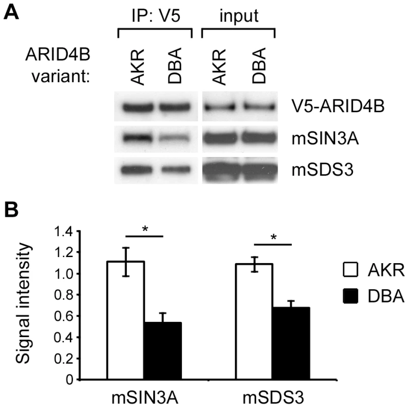
Western blots (A) show equivalent input of V5-ARID4B, mSIN3A, mSDS3, and equivalent pull-down of the two variants using an anti-V5 antibody. Decreased binding of the DBA variant of ARID4B to mSIN3A and mSDS3 was observed relative to the AKR variant. Experiment was performed in triplicate and densitometry analysis (B) revealed a 51% reduction in mSIN3A binding (p = .037) and a 37% reduction in mSDS3 binding (p = .026) for the DBA variant relative to AKR. Statistical significance was determined using paired t-tests. *, p<.05. ARID4B binds the breast cancer metastasis suppressor BRMS1
Breast cancer metastasis suppressor 1 (BRMS1) belongs to the same family of proteins as mSDS3 and is known to associate with the mSIN3A complex as well as ARID4A [13]. Because ARID4B is also known to bind mSIN3A, mSDS3, and ARID4A [14], we postulated that ARID4B might physically bind BRMS1. Proteomics screens to identify BRMS1 interacting proteins also support this association: in a yeast two-hybrid screen for proteins binding full-length BRMS1, ARID4B was the number one hit identified, and ARID4A and mSDS3 were also detected (unpublished data). In a separate screen, mass spectrometry was performed to identify mSIN3A binding proteins in MCF10A human breast epithelial cells. Peptides representing endogenous ARID4B and BRMS1 were detected, providing further evidence for this interaction and demonstrating that it is not simply an artifact of supraphysiologic expression in transfected cells (Douglas Hurst; personal communication). To validate this interaction we performed co-IPs using lysates from 293 cells transiently transfected with the FLAG-tagged AKR or DBA variants of ARID4B along with either HA - or myc-tagged BRMS1. HA-BRMS1 was readily detected following pull-down of ARID4B using an anti-FLAG antibody (Figure 6A). Likewise, ARID4B was efficiently co-precipitated with myc-BRMS1 (Figure 6B). Unlike the associations with mSIN3A and mSDS3 however, the AKR and DBA variants of ARID4B did not exhibit differential binding to BRMS1. One possible explanation for this observation is that BRMS1 binds to a different region of ARID4B than the polymorphic C-terminal domain that mediates binding to mSIN3A.
Fig. 6. ARID4B binds BRMS1. 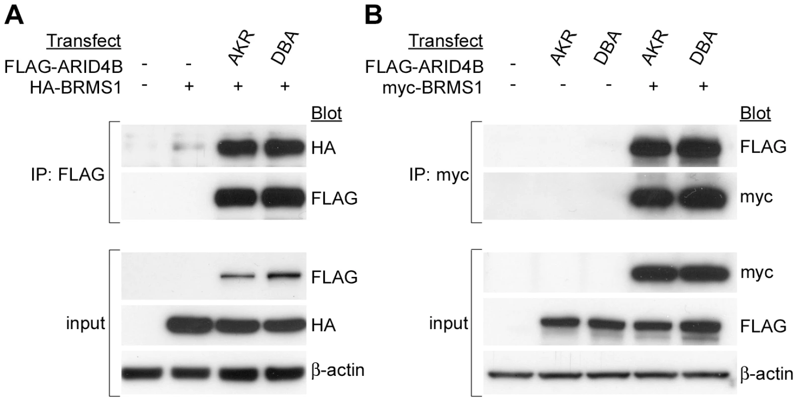
HA-BRMS1 was coprecipitated with FLAG-ARID4B (A) and FLAG-ARID4B was coprecipitated with myc-BRMS1 (B). No difference in binding to BRMS1 was observed between the AKR and DBA alleles of ARID4B. Arid4b regulates the metastasis-associated TPX2 gene network
Because little is known about the specific cellular processes regulated by Arid4b that might influence the metastatic phenotype, we performed expression microarray analysis on the 6DT1 cell lines stably expressing Arid4b shRNAs to identify genes that are differentially expressed as a function of Arid4b levels. Based on the western blot densitometry shown in Figure 4A–4B cell lines expressing hairpins H3 and H4 were chosen to represent the Arid4b knockdown cohort, and the control cohort consisted of untreated 6DT1 cells and lines expressing the scrambled control shRNA or hairpin H5. We detected 2,048 unique genes whose expression was significantly different (p<0.05, ANOVA) between the two groups and those with the greatest fold change are summarized in Table 1. While the most highly upregulated genes function in pathways with diverse biological roles, it was noted that among the most downregulated genes were multiple factors associated with centromeres (Cenpi, Cenpq), microtubule and spindle dynamics (Kif2c, Kif4a, Sass6), and cell cycle regulation (Ccne, Cdc25c). Consistent with this observation were the results of pathway analysis conducted to identify biological functions impacted as a consequence of Arid4b knockdown (Table 2). The most differentially regulated processes based on gene ontology were checkpoint control and DNA repair, and processes related to centrosome, centriole, and chromosome dynamics.
Tab. 1. Genes most highly up- or down-regulated following <i>Arid4b</i> knockdown. 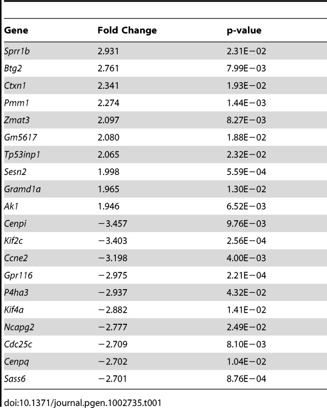
Tab. 2. Cellular processes regulated by Arid4b knockdown. 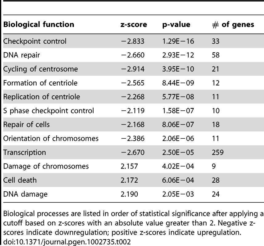
Biological processes are listed in order of statistical significance after applying a cutoff based on z-scores with an absolute value greater than 2. Negative z-scores indicate downregulation; positive z-scores indicate upregulation. In examining the microarray data we noticed a striking overlap between genes downregulated in the Arid4b knockdown lines and components of the TPX2 gene network. This transcriptional network was recently identified based on expression profiling of three mouse data sets and two human breast cancer data sets [6]. The TPX2 network is tumor cell-autonomous and conserved across species, its activation is predictive of reduced distant metastasis-free survival (DMFS) in ER-positive patients, and the nine common hub genes in the TPX2 signature (TPX2, BUB1, UBE2C, CDC20, CCNB2, KIF2C, BUB1B, CEP55, CENPA) that were conserved across all five data sets consist primarily of genes involved in microtubule and mitotic spindle function. To determine how Arid4b levels influence the activation state of the TPX2 network, the fold changes of the 311 TPX2 network genes were examined in the Arid4b knockdown lines. Compared to control cell lines, 119 network genes were significantly downregulated (p<0.05) including Tpx2 itself and the other eight common hub genes, versus only 5 network genes upregulated (Figure 7; high resolution available as Figure S3). The downregulation of this gene network concomitant with the inhibition of metastasis observed in the Arid4b knockdown lines provides further support for the role of the TPX2 network in metastatic susceptibility and suggests that a significant portion of this network may be regulated by Arid4b.
Fig. 7. Expression of Tpx2 network genes in Arid4b knockdown cell lines. 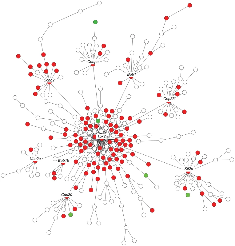
The nine labeled hub genes were conserved across mouse and human breast cancer data sets. Red indicates statistically significant (p<.05) downregulation; green indicates upregulation. High Arid4b expression predicts poor clinical outcome
Because Arid4b was identified as a candidate gene in part based on differential expression between high and low metastatic strains of mice in the AKXD panel, and because Arid4b expression levels were associated with tumor growth and metastasis in mice as well as the activity of the metastasis-associated TPX2 network, we tested whether ARID4B expression alone correlated with human patient outcomes. A search of publically available breast cancer microarray data sets using Oncomine (Compendia Bioscience, Ann Arbor, MI) revealed that ARID4B expression was 2.3-fold higher in 40 ductal breast carcinoma samples compared to 7 normal breast tissue samples in the Richardson study [15], confirming that high ARID4B expression is clinically associated with breast cancer (Figure S4). Analysis of a pooled breast cancer dataset using GOBO (http://co.bmc.lu.se/gobo/) [16] showed that among the subgroup of patients with ER-positive tumors, the cohort with high expression of ARID4B had significantly reduced DMFS compared to the low or median ARID4B cohorts (Figure 8). Because this association was significant among patients with ER-positive tumors who were lymph node negative at the time of diagnosis (Figure 8A), this finding indicated that ARID4B expression level is predictive of patient progression to metastatic disease. As determined by multivariate analysis (Figure 8B), the hazard ratio compared to the high ARID4B tercile was 0.54 for middle ARID4B (95% C.I. = 0.33–0.89; p = .015) and 0.42 for the low ARID4B tercile (95% C.I. = 0.26–0.70; p = 7.51×10∧−4), indicating that patients with tumors expressing high levels of ARID4B are approximately twice as likely to develop metastatic disease. The association of ARID4B with reduced DMFS was also highly significant among ER-positive patients not receiving adjuvant therapy (Figure 8C–8D), indicating that ARID4B expression level plays a significant role in the natural metastatic progression of ER-positive breast cancer in human patients and its relevance is not confined solely to our mouse model systems.
Fig. 8. High expression of ARID4B is associated with poor clinical outcome. 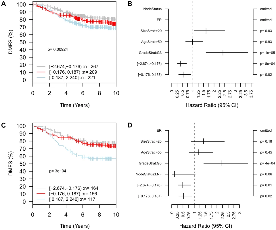
In patients with ER-positive tumors who were node negative at diagnosis (A) distant metastasis-free survival (DMFS) was significantly lower (p = .009) in patients with high expression (blue) compared to middle (red) or low (gray) expression of ARID4B, and multivariate analysis of 440 cases (B) was performed to determine metastatic progression hazard ratios of 0.54 and 0.42 for median and low ARID4B terciles, respectively, compared to the high ARID4B tercile. The association of high ARID4B with poor DMFS was also highly significant (p = 3.05×10∧−4) among ER-positive patients in the absence of adjuvant therapy (C) with similar hazard ratios (D) of 0.53 and 0.49 for middle and low ARID4B groups compared to high ARID4B. Discussion
Arid4b was identified as a candidate gene of interest through linkage and expression correlation analyses, and the in vitro and in vivo data presented here provide the first direct evidence of a causal role of Arid4b in mammary tumor progression and metastasis. The initial QTL analysis revealed association with Arid4b on proximal chromosome 13, and Arid4b was among the most highly correlated genes with the most significant p values in the subsequent eQTL analysis in the AKXD recombinant inbred panel. It was noted that although AKR/J is the more highly metastatic of the two parental strains, progression was associated with the DBA/2J allele, suggesting that the metastasis promoting influence of Arid4b is likely masked by other suppressive factors in a pure DBA/2J background. Although the AKXD recombinant inbred panel lacks the power to detect these epistatic interactions, ongoing experiments using the latest generation of recombinant inbred mice including the Collaborative Cross [17], [18] will enable higher resolution QTL mapping and more robust systems genetics analyses going forward.
Mouse Arid4b encodes a protein of 1314 amino acids that shares 89% identity and 95% similarity to the 1312 amino acid human protein. Alternate nomenclature includes breast cancer-associated antigen 1 (BRCAA1), retinoblastoma-binding protein-1-like protein-1 (RBP1L1), and mSIN3A-associated protein of 180 kDa (SAP180). Indeed, there are multiple lines of evidence implicating Arid4b in breast cancer. A ten amino acid peptide was found to represent an antigen epitope expressed in 65% of breast cancer specimens and was significantly upregulated in the sera of breast cancer patients compared to healthy donors [19]. ARID4B was also found to associate with the mSIN3A HDAC complex [5], which is in turn known to be bound by the breast cancer associated tumor suppressor ING1 [20], [21], the well-characterized breast cancer metastasis suppressor BRMS1 [13], and the ARID family homolog ARID4A/RBP1 [22], which has also been identified as a breast cancer associated antigen [23].
Ectopic expression of Arid4b at a physiologically relevant two - to three-fold increased level resulted in a 3-fold increase in orthotopic tumor mass relative to controls for Met-1 cells expressing the DBA allele, while the AKR allele induced 1.9-fold larger tumors versus control cells. To our knowledge, this is the first direct evidence that Arid4b upregulation promotes tumor growth. Although Met-1 orthotopic tumors do not readily metastasize in our experience, transwell assays in vitro demonstrated that upregulation of either allele of Arid4b increased tumor cell migration and invasion, consistent with a role of Arid4b in metastatic progression, and cells expressing the DBA allele were significantly more migratory and invasive than cells expressing the AKR allele. While stable upregulation of Arid4b did not induce Met-1 cells to metastasize with any greater frequency, stable knockdown of Arid4b in the highly metastatic 6DT1 cell line did cause a dramatic reduction in pulmonary metastases, raising the possibility that ARID4B may represent a novel therapeutic target. Taken together, the results of the orthotopic implantation and transwell assays are broadly consistent with our genetic linkage and expression correlation analyses that showed an association of the DBA haplotype on chromosome 13 with metastatic progression in the MMTV-PyMT×AKXD mice, and validate a functional role of Arid4b polymorphism in modulating the breast cancer phenotype.
While the molecular mechanisms of Arid4b are incompletely understood, an examination of its sequence and conserved domains provides further insight into its potential functions. Arid4b contains a nuclear localization signal (NLS) towards the C-terminus as well as conserved Tudor, RBB1NT, ARID/BRIGHT, and Chromo domains in the N-terminal half of the protein. The ARID domain mediates binding to DNA, although the affinity for AT-rich sequences varies among members of the Arid superfamily [24], [25]. The RBB1NT domain is present in many Rb binding proteins including ARID4A, although it is noteworthy that unlike ARID4A, ARID4B does not contain the LCXCE motif necessary for RB binding [26], and no interaction was observed when we attempted to co-IP ARID4B with RB (data not shown); therefore, the function of the RBB1NT domain of ARID4B remains uncertain. Tudor domains are present in many RNA binding proteins [27] and also bind methylated lysine residues on histone tails [28]. Chromo domain-containing proteins have also been shown to bind methylated lysines and mediate the recruitment of chromatin modifying complexes [29]. Because mSIN3A itself lacks intrinsic DNA binding capability, targeting of mSIN3A-associated HDAC activity depends on interactions with other transcription factors including Mad1 and KLF repressors among others [30]. The presence of putative DNA and histone binding domains in the N-terminal half of ARID4B suggest that its influence on mammary tumor progression involves directing the HDAC activity of mSIN3A complexes to chromatin. This is supported by our observations that the high and low metastatic alleles of ARID4B have a dense cluster of amino acid polymorphisms in the C-terminal domain and bind with different affinities to mSIN3A and mSDS3, though the biochemical significance of this observation remains to be determined. Diminished expression levels or binding affinity of ARID4B may allow mSIN3A to be bound by other proteins with different DNA sequence specificity, perhaps not resulting in a global change in the abundance of any one particular histone mark but rather altering the expression of different subsets of genes.
It is noteworthy that the pro-metastatic ARID4B and the metastasis suppressive BRMS1 bind each other and also to the mSIN3A complex in vitro. This observation reinforces the significance of the mSIN3A complex in metastatic progression, and it is tempting to speculate that an HDAC complex may be caught in a molecular tug-of-war between these two metastasis modifier genes. However, the mSIN3A complex is modular in nature and interacts with a great variety of transcriptional regulators [30], [31]. Many different complexes exist, and their precise composition and function within the context of breast cancer are not well understood. Further studies will be necessary to define a role, if any, for the ARID4B-BRMS1 interaction in human disease.
While ARID4B expression was not a significant predictor of DMFS across all patients in a meta-analysis of an 1,881 sample data set, statistical significance emerged when patients were stratified based on ER status. The observation that ARID4B is predictive of metastatic progression only in ER+ patients is consistent with the identification of Arid4b as a candidate gene in the context of the MMTV-PyMT mouse model system, in which tumors arise from a predominantly ER+ luminal epithelial cell population [32]. Loss of ER and PR is detected during progression to late carcinomas, however in a systematic analysis of gene expression profiles these late PyMT tumors clustered most closely with human luminal tumors [33], which are ER+. Also consistent with ARID4B promoting metastatic progression of ER+ tumors is our observation that Arid4b knockdown caused a significant downregulation of the core components of the Tpx2 gene network. The TPX2 signature was tumor cell autonomous and predictive of DMFS only in those patients who were ER+ at diagnosis, and was distinct from a CD53 network that was associated with ER-negative stromal components [6]. Polymorphisms in several other tumor cell autonomous metastasis susceptibility genes identified in our laboratory including Sipa1, Rrp1b, and Brd4 are prognostic only in ER+ patients [9], [34]–[37], and stable expression of Brd4 can also differentially regulate the Tpx2 network [6]. The association of multiple metastasis susceptibility genes with a transcriptional network comprising many cell cycle and mitotic spindle checkpoint regulatory genes highlights the possibility that these cellular functions are critical determinants of metastatic efficiency. Further experiments are underway in our laboratory to determine whether upregulation of the TPX2 network is causative in promoting metastasis.
Materials and Methods
Identification of Arid4b germline polymorphisms
Using genomic DNA from AKR/J and DBA/2J mice as templates, PCR was performed to amplify the protein coding region, exons 2 through 24 (Table S1). PCR Products were subjected to agarose gel electrophoresis, bands isolated using the QIAquick Gel Extraction kit (Qiagen) according to manufacturer's recommendations, and used as templates for sequencing. All sequencing runs were performed by the DNA Sequencing and Gene Expression Core, NCI, Bethesda, MD. Genomic sequences for the AKR and DBA alleles were aligned using pairwise BLAST [38] and non-synonymous polymorphisms verified by manual comparison of chromatograms using Chromas software (Technelysium).
Expression vectors
V5-tagged AKR and DBA alleles of Arid4b were generated using long range PCR with forward primer 5′-AACAAAGGTGCAGGTGAAGC-3′ and reverse primer 5′-CCTGCACTCAACTGACATTCCATTC-3′ to amplify Arid4b, and PCR products were cloned into pcDNA3.1/V5-His-TOPO (Invitrogen). FLAG-tagged Arid4b vectors were constructed by the Protein Expression Laboratory, SAIC-Frederick, Inc. using Gateway technology (Invitrogen). Briefly, the AKR or DBA allele of Arid4b was PCR amplified and cloned into entry vector pDonr-253, then subcloned by Gateway LR recombination into pDest-737 to generate an expression construct with CMV promoter and N-terminal 3xFLAG tag. Full-length BRMS1 was epitope tagged at the N-terminus by PCR with the HA or myc tag sequence incorporated into the forward primer and cloned into pCMV or pcDNA3-hygro (Invitrogen), respectively. Correct sequences of all vectors were confirmed prior to use.
Cell culture and stable cell lines
Met-1 cells [10] were a gift from Dr. Robert Cardiff (University of California, Davis, CA). 6DT1 cells [11] were a gift from Dr. Lalage Wakefield (NCI, NIH, Bethesda, MD). HEK293 cells were purchased from ATCC (Manassas, VA). Cell lines were maintained in DMEM supplemented with 10% FBS, 2 mM L-glutamine, penicillin and streptomycin. Cells were confirmed to be free of mycoplasma contamination using the MycoAlert detection kit (Lonza).
Met-1 cells seeded onto 10 cm tissue culture plates were co-transfected with 6 µg of the appropriate V5-tagged Arid4b construct described above, or pcDNA3.1/V5-His-TOPO/lacZ (Invitrogen) as a control vector, plus 600 ng of pSuper.Retro.Puro (Oligoengine) as a selectable marker, using FuGENE 6 transfection reagent. Cells were selected using 1 mg/ml G418 plus 4 µg/ml puromycin and clones derived by limiting dilution. Stable upregulation of Arid4b was verified by performing QRT-PCR using forward primer 5′ - GGTGAGTGGGAGCTGGTCTA-3′ and reverse primer 5′ - ATAAAGGGCCCACTGAAGGT-3′, and western blotting for endogenous and ectopically expressed ARID4B as described below. 6DT1 cells were transduced with one of five different lentiviral shRNAs targeting Arid4b (RMM4534-NM_194262, Open Biosystems) or a scrambled control shRNA in the same pLKO.1 vector. Stable cells were selected using 10 µg/ml puromycin and pooled clones were analyzed for Arid4b knockdown by western blot.
Animal studies
Met-1 or 6DT1 stable lines were orthotopically implanted into the fourth mammary fat pad of six week old female NU/J or FVB/NJ mice using 105 cells suspended in 100 µl of PBS per animal. Primary tumors and lungs were harvested 28 days later. All experiments were performed according to the National Cancer Institute Animal Care and Use Committee guidelines.
Migration and invasion assays
Met-1 cells stably expressing Arid4b or lacZ were seeded at 75,000 cells per well into invasion chambers coated with Matrigel basement membrane matrix (534480, BD Biosciences) or control chambers lacking Matrigel (354578, BD Biosciences). After 24 hours, cells were fixed in 100% methanol, stained with crystal violet, and mounted onto glass slides using mineral oil. Cells were visualized at 400× magnification and five fields were counted for each of three experiments.
Co-immunoprecipitations
HEK293 cells were transfected with the V5 - or FLAG-tagged AKR or DBA allele of Arid4b, with or without HA-BRMS1 or myc-BRMS1 where appropriate, using FuGENE 6 transfection reagent. After 30 hours, cells were harvested in mild IP lysis buffer (25 mM Tris-HCl pH 7.4, 150 mM NaCl, 1 mM EDTA, 1% NP-40, 5% glycerol) supplemented with protease inhibitors (11836170001, Roche) and phosphatase inhibitors (P-5726, Sigma). Protein samples were quantitated using Bradford assays. Gammabind G Sepharose beads (17088501, GE Healthcare) were washed twice in NET buffer (50 mM Tris pH 8.0, 150 mM NaCl, 5 mM EDTA, 1% NP-40, 0.5% BSA, 0.04% sodium azide) supplemented with protease and phosphatase inhibitors, and resuspended to form a 50% bead slurry. Lysates were precleared by adding 40 µl of bead slurry and rotating for 30 minutes at 4°C. Samples were centrifuged at 10,000 rpm for 1 minute at 4°C and pre-cleared supernatant transferred to a fresh tube. Anti-V5, anti-FLAG, or anti-myc tag antibodies was added to a final concentration of 1.0 µg/ml and samples rotated for 1 hour at 4°C, then 50 µl of bead slurry was added and co-IPs performed overnight at 4°C. Beads were then washed four times with NET buffer and resuspended in SDS-PAGE sample buffer.
Western blots and densitometry
NuPAGE precast gels and buffers (Invitrogen) were used according to manufacturers recommendations and gels were transferred onto Immobilon-P (Millipore). Membranes were blocked in TBS with 0.5% Tween-20 (TBST) plus 5% nonfat dry milk for 1 hour at room temperature then incubated with the primary antibody diluted in blocking buffer overnight at 4°C. The following primary antibodies and concentrations were used: anti-Arid4b (1∶3,000; A302-233A, Bethyl Laboratories), anti-V5 (1∶5,000; 37–7500, Invitrogen), anti-mSIN3A (1∶1,000; sc-994, Santa Cruz), anti-mSDS3 (1∶2,000; A300-235A, Bethyl Labs), anti-β-actin (1∶10,000; ab6276, Abcam), anti-FLAG (1∶3,000; F-3165, Sigma), anti-HA (1∶5,000; 11867423001, Roche), anti-myc tag (1∶2,000; 2276, Cell Signaling). After three washes in TBST, membranes were incubated with one of the following horseradish peroxidase conjugated secondary antibodies diluted in TBST plus 0.5% milk for 1 hour at room temperature: anti-mouse (1∶5,000; NA931V, GE Healthcare), anti-rabbit (1∶10,000; sc-2004, Santa Cruz), anti-goat (1∶10,000; sc-2304, Santa Cruz). Membranes were washed an additional three times in TBST and proteins detected using the Amersham ECL Plus system (RPN2132, GE Healthcare) and Amersham Hyperfilm ECL (28906837, GE Healthcare) according to manufacturer's recommendations. Densitometry data were collected and analyzed using a ChemiDoc-It Imaging System and VisionWorksLS software (UVP).
Expression profiling and network analysis
Total RNA was isolated from pooled clones of 6DT1 Arid4b knockdown cell lines using RNeasy kits (Qiagen) and then arrayed on Affymetrix GeneChip Mouse Gene 1.0 ST arrays by the Microarray Core in the NCI Laboratory of Molecular Technology. Expression data were normalized using Partek Genomics Suite to identify genes whose expression was significantly different (p<.05) between the Arid4b normal cohort (untreated, scrambled control, and H5 lines) and the Arid4b knockdown cohort (lines H3 and H4). The gene list and expression values were then analyzed using Ingenuity Pathways Analysis (Ingenuity Systems, www.ingenuity.com) to identify differentially regulated signaling pathways and biological functions. Expression of the Tpx2 transcriptional network was visualized and figure generated using Cytoscape software [39]. Microarray data are available through the Gene Expression Omnibus under accession number GSE35731.
Supporting Information
Zdroje
1. American Cancer Society 2012 Cancer Facts and Figures 2012 Atlanta American Cancer Society
2. LifstedTLe VoyerTWilliamsMMullerWKlein-SzantoA 1998 Identification of inbred mouse strains harboring genetic modifiers of mammary tumor age of onset and metastatic progression. Int J Cancer 77 640 644
3. HunterKWBromanKWVoyerTLLukesLCozmaD 2001 Predisposition to efficient mammary tumor metastatic progression is linked to the breast cancer metastasis suppressor gene Brms1. Cancer Res 61 8866 8872
4. LukesLCrawfordNPWalkerRHunterKW 2009 The origins of breast cancer prognostic gene expression profiles. Cancer Res 69 310 318
5. FleischerTCYunUJAyerDE 2003 Identification and characterization of three new components of the mSin3A corepressor complex. Mol Cell Biol 23 3456 3467
6. HuYWuGRuschMLukesLBuetowKH 2012 Integrated cross-species transcriptional network analysis of metastatic susceptibility. Proc Natl Acad Sci USA 109 8 3184 3189
7. YangHCrawfordNLukesLFinneyRLancasterM 2005 Metastasis predictive signature profiles pre-exist in normal tissues. Clin Exp Metastasis 22 593 603
8. WuCCHuangHCJuanHFChenST 2004 GeneNetwork: an interactive tool for reconstruction of genetic networks using microarray data. Bioinformatics 20 3691 3693
9. ParkYGZhaoXLesueurFLowyDRLancasterM 2005 Sipa1 is a candidate for underlying the metastasis efficiency modifier locus Mtes1. Nat Genet 37 1055 1062
10. BorowskyADNambaRYoungLJHunterKWHodgsonJG 2005 Syngeneic mouse mammary carcinoma cell lines: two closely related cell lines with divergent metastatic behavior. Clin Exp Metastasis 22 47 59
11. PeiXFNobleMSDavoliMARosfjordETilliMT 2004 Explant-cell culture of primary mammary tumors from MMTV-c-Myc transgenic mice. In Vitro Cell Dev Biol Anim 40 14 21
12. AllandLDavidGShen-LiHPotesJMuhleR 2002 Identification of mammalian Sds3 as an integral component of the Sin3/histone deacetylase corepressor complex. Mol Cell Biol 22 2743 2750
13. MeehanWJSamantRSHopperJECarrozzaMJShevdeLA 2004 Breast cancer metastasis suppressor 1 (BRMS1) forms complexes with retinoblastoma-binding protein 1 (RBP1) and the mSin3 histone deacetylase complex and represses transcription. J Biol Chem 279 1562 1569
14. WuMYTsaiTFBeaudetAL 2006 Deficiency of Rbbp1/Arid4a and Rbbp1l1/Arid4b alters epigenetic modifications and suppresses an imprinting defect in the PWS/AS domain. Genes Dev 20 2859 2870
15. RichardsonALWangZCDe NicoloALuXBrownM 2006 X chromosomal abnormalities in basal-like human breast cancer. Cancer Cell 9 121 132
16. RingnerMFredlundEHakkinenJBorgAStaafJ GOBO: Gene Expression-Based Outcome for Breast Cancer Online. PLoS One 6 e17911 doi:10.1371/journal.pone.0017911
17. AylorDLValdarWFoulds-MathesWBuusRJVerdugoRA Genetic analysis of complex traits in the emerging Collaborative Cross. Genome Res 21 1213 1222
18. ThreadgillDWChurchillGA Ten years of the collaborative cross. Genetics 190 291 294
19. CuiDJinGGaoTSunTTianF 2004 Characterization of BRCAA1 and its novel antigen epitope identification. Cancer Epidemiol Biomarkers Prev 13 1136 1145
20. ToyamaTIwaseHWatsonPMuzikHSaettlerE 1999 Suppression of ING1 expression in sporadic breast cancer. Oncogene 18 5187 5193
21. SkowyraDZeremskiMNeznanovNLiMChoiY 2001 Differential association of products of alternative transcripts of the candidate tumor suppressor ING1 with the mSin3/HDAC1 transcriptional corepressor complex. J Biol Chem 276 8734 8739
22. LaiAKennedyBKBarbieDABertosNRYangXJ 2001 RBP1 recruits the mSIN3-histone deacetylase complex to the pocket of retinoblastoma tumor suppressor family proteins found in limited discrete regions of the nucleus at growth arrest. Mol Cell Biol 21 2918 2932
23. CaoJGaoTGiulianoAEIrieRF 1999 Recognition of an epitope of a breast cancer antigen by human antibody. Breast Cancer Res Treat 53 279 290
24. WilskerDPatsialouADallasPBMoranE 2002 ARID proteins: a diverse family of DNA binding proteins implicated in the control of cell growth, differentiation, and development. Cell Growth Differ 13 95 106
25. DallasPBPacchioneSWilskerDBowrinVKobayashiR 2000 The human SWI-SNF complex protein p270 is an ARID family member with non-sequence-specific DNA binding activity. Mol Cell Biol 20 3137 3146
26. Defeo-JonesDHuangPSJonesREHaskellKMVuocoloGA 1991 Cloning of cDNAs for cellular proteins that bind to the retinoblastoma gene product. Nature 352 251 254
27. PontingCP 1997 Tudor domains in proteins that interact with RNA. Trends Biochem Sci 22 51 52
28. BotuyanMVLeeJWardIMKimJEThompsonJR 2006 Structural basis for the methylation state-specific recognition of histone H4-K20 by 53BP1 and Crb2 in DNA repair. Cell 127 1361 1373
29. EissenbergJC 2001 Molecular biology of the chromo domain: an ancient chromatin module comes of age. Gene 275 19 29
30. GrzendaALomberkGZhangJSUrrutiaR 2009 Sin3: master scaffold and transcriptional corepressor. Biochim Biophys Acta 1789 443 450
31. SilversteinRAEkwallK 2005 Sin3: a flexible regulator of global gene expression and genome stability. Curr Genet 47 1 17
32. LinEYJonesJGLiPZhuLWhitneyKD 2003 Progression to malignancy in the polyoma middle T oncoprotein mouse breast cancer model provides a reliable model for human diseases. Am J Pathol 163 2113 2126
33. HerschkowitzJISiminKWeigmanVJMikaelianIUsaryJ 2007 Identification of conserved gene expression features between murine mammary carcinoma models and human breast tumors. Genome Biol 8 R76
34. CrawfordNPQianXZiogasAPapageorgeAGBoersmaBJ 2007 Rrp1b, a new candidate susceptibility gene for breast cancer progression and metastasis. PLoS Genet 3 e214 doi:10.1371/journal.pgen.0030214
35. AlsarrajJWalkerRCWebsterJDGeigerTRCrawfordNP 2011 Deletion of the Proline-Rich Region of the Murine Metastasis Susceptibility Gene Brd4 Promotes Epithelial-to-Mesenchymal Transition - and Stem Cell-Like Conversion. Cancer Res 71 3121 3131
36. CrawfordNPWalkerRCLukesLOfficewalaJSWilliamsR 2008 The Diasporin Pathway: a tumor progression-related transcriptional network that predicts breast cancer survival. Clin Exp Metastasis
37. HsiehSMLookMPSieuwertsAMFoekensJAHunterKW 2009 Distinct inherited metastasis susceptibility exists for different breast cancer subtypes: a prognosis study. Breast Cancer Res 11 R75
38. ZhangZSchwartzSWagnerLMillerW 2000 A greedy algorithm for aligning DNA sequences. J Comput Biol 7 203 214
39. ShannonPMarkielAOzierOBaligaNSWangJT 2003 Cytoscape: a software environment for integrated models of biomolecular interaction networks. Genome Res 13 2498 2504
Štítky
Genetika Reprodukční medicína
Článek Functional Centromeres Determine the Activation Time of Pericentric Origins of DNA Replication inČlánek Dynamic Deposition of Histone Variant H3.3 Accompanies Developmental Remodeling of the TranscriptomeČlánek Integrin α PAT-2/CDC-42 Signaling Is Required for Muscle-Mediated Clearance of Apoptotic Cells inČlánek Prdm5 Regulates Collagen Gene Transcription by Association with RNA Polymerase II in Developing BoneČlánek Acquisition Order of Ras and p53 Gene Alterations Defines Distinct Adrenocortical Tumor Phenotypes
Článek vyšel v časopisePLOS Genetics
Nejčtenější tento týden
2012 Číslo 5
-
Všechny články tohoto čísla
- Slowing Replication in Preparation for Reduction
- Chromosome Pairing: A Hidden Treasure No More
- Loss of Imprinting Differentially Affects REM/NREM Sleep and Cognition in Mice
- Six Novel Susceptibility Loci for Early-Onset Androgenetic Alopecia and Their Unexpected Association with Common Diseases
- Regulation by the Noncoding RNA
- UDP-Galactose 4′-Epimerase Activities toward UDP-Gal and UDP-GalNAc Play Different Roles in the Development of
- Deletion of PTH Rescues Skeletal Abnormalities and High Osteopontin Levels in Mice
- Karyotypic Determinants of Chromosome Instability in Aneuploid Budding Yeast
- Genome-Wide Copy Number Analysis Uncovers a New HSCR Gene:
- MicroRNA-277 Modulates the Neurodegeneration Caused by Fragile X Premutation rCGG Repeats
- Functional Centromeres Determine the Activation Time of Pericentric Origins of DNA Replication in
- Dynamic Deposition of Histone Variant H3.3 Accompanies Developmental Remodeling of the Transcriptome
- Scientist Citizen: An Interview with Bruce Alberts
- YY1 Regulates Melanocyte Development and Function by Cooperating with MITF
- Congenital Heart Disease–Causing Gata4 Mutation Displays Functional Deficits
- Recombination Drives Vertebrate Genome Contraction
- KATNAL1 Regulation of Sertoli Cell Microtubule Dynamics Is Essential for Spermiogenesis and Male Fertility
- Re-Patterning Sleep Architecture in through Gustatory Perception and Nutritional Quality
- Using Whole-Genome Sequence Data to Predict Quantitative Trait Phenotypes in
- Genome-Wide Analysis of GLD-1–Mediated mRNA Regulation Suggests a Role in mRNA Storage
- Meiotic Chromosome Pairing Is Promoted by Telomere-Led Chromosome Movements Independent of Bouquet Formation
- LINT, a Novel dL(3)mbt-Containing Complex, Represses Malignant Brain Tumour Signature Genes
- The H3K27 Demethylase UTX-1 Is Essential for Normal Development, Independent of Its Enzymatic Activity
- Suppresses Senescence Programs and Thereby Accelerates and Maintains Mutant -Induced Lung Tumorigenesis
- Genome-Wide Association of Pericardial Fat Identifies a Unique Locus for Ectopic Fat
- An Essential Role for Katanin p80 and Microtubule Severing in Male Gamete Production
- Identification of Genes That Promote or Antagonize Somatic Homolog Pairing Using a High-Throughput FISH–Based Screen
- Principles of Carbon Catabolite Repression in the Rice Blast Fungus: Tps1, Nmr1-3, and a MATE–Family Pump Regulate Glucose Metabolism during Infection
- Integrin α PAT-2/CDC-42 Signaling Is Required for Muscle-Mediated Clearance of Apoptotic Cells in
- Histone H3 Localizes to the Centromeric DNA in Budding Yeast
- Collapse of Telomere Homeostasis in Hematopoietic Cells Caused by Heterozygous Mutations in Telomerase Genes
- Hypersensitive to Red and Blue 1 and Its Modification by Protein Phosphatase 7 Are Implicated in the Control of Arabidopsis Stomatal Aperture
- Extent, Causes, and Consequences of Small RNA Expression Variation in Human Adipose Tissue
- TBC-8, a Putative RAB-2 GAP, Regulates Dense Core Vesicle Maturation in
- Regulating Repression: Roles for the Sir4 N-Terminus in Linker DNA Protection and Stabilization of Epigenetic States
- Common Genetic Determinants of Intraocular Pressure and Primary Open-Angle Glaucoma
- Prdm5 Regulates Collagen Gene Transcription by Association with RNA Polymerase II in Developing Bone
- Fitness Landscape Transformation through a Single Amino Acid Change in the Rho Terminator
- Repeated, Selection-Driven Genome Reduction of Accessory Genes in Experimental Populations
- Allelic Variation and Differential Expression of the mSIN3A Histone Deacetylase Complex Gene Promote Mammary Tumor Growth and Metastasis
- DNA Demethylation and USF Regulate the Meiosis-Specific Expression of the Mouse
- Knowledge-Driven Analysis Identifies a Gene–Gene Interaction Affecting High-Density Lipoprotein Cholesterol Levels in Multi-Ethnic Populations
- A Duplication CNV That Conveys Traits Reciprocal to Metabolic Syndrome and Protects against Diet-Induced Obesity in Mice and Men
- EMT Inducers Catalyze Malignant Transformation of Mammary Epithelial Cells and Drive Tumorigenesis towards Claudin-Low Tumors in Transgenic Mice
- Inactivation of a Novel FGF23 Regulator, FAM20C, Leads to Hypophosphatemic Rickets in Mice
- Genome-Wide Association for Abdominal Subcutaneous and Visceral Adipose Reveals a Novel Locus for Visceral Fat in Women
- Stratifying Type 2 Diabetes Cases by BMI Identifies Genetic Risk Variants in and Enrichment for Risk Variants in Lean Compared to Obese Cases
- New Insight into the History of Domesticated Apple: Secondary Contribution of the European Wild Apple to the Genome of Cultivated Varieties
- Activated Cdc42 Kinase Has an Anti-Apoptotic Function
- The Region Is Critical for Birth Defects and Electrocardiographic Dysfunctions Observed in a Down Syndrome Mouse Model
- COP9 Signalosome Integrity Plays Major Roles for Hyphal Growth, Conidial Development, and Circadian Function
- Bmps and Id2a Act Upstream of Twist1 To Restrict Ectomesenchyme Potential of the Cranial Neural Crest
- Psip1/Ledgf p52 Binds Methylated Histone H3K36 and Splicing Factors and Contributes to the Regulation of Alternative Splicing
- The Number of X Chromosomes Causes Sex Differences in Adiposity in Mice
- Target Gene Analysis by Microarrays and Chromatin Immunoprecipitation Identifies HEY Proteins as Highly Redundant bHLH Repressors
- Acquisition Order of Ras and p53 Gene Alterations Defines Distinct Adrenocortical Tumor Phenotypes
- ELK1 Uses Different DNA Binding Modes to Regulate Functionally Distinct Classes of Target Genes
- Histone H1 Depletion Impairs Embryonic Stem Cell Differentiation
- IDN2 and Its Paralogs Form a Complex Required for RNA–Directed DNA Methylation
- Separation of DNA Replication from the Assembly of Break-Competent Meiotic Chromosomes
- Genomic Hypomethylation in the Human Germline Associates with Selective Structural Mutability in the Human Genome
- PLOS Genetics
- Archiv čísel
- Aktuální číslo
- Informace o časopisu
Nejčtenější v tomto čísle- Inactivation of a Novel FGF23 Regulator, FAM20C, Leads to Hypophosphatemic Rickets in Mice
- Genome-Wide Association of Pericardial Fat Identifies a Unique Locus for Ectopic Fat
- Slowing Replication in Preparation for Reduction
- An Essential Role for Katanin p80 and Microtubule Severing in Male Gamete Production
Kurzy
Zvyšte si kvalifikaci online z pohodlí domova
Současné možnosti léčby obezity
nový kurzAutoři: MUDr. Martin Hrubý
Všechny kurzyPřihlášení#ADS_BOTTOM_SCRIPTS#Zapomenuté hesloZadejte e-mailovou adresu, se kterou jste vytvářel(a) účet, budou Vám na ni zaslány informace k nastavení nového hesla.
- Vzdělávání


