Antagonism of miR-328 Increases the Antimicrobial Function of Macrophages and Neutrophils and Rapid Clearance of Non-typeable (NTHi) from Infected Lung
MicroRNAs regulate pathogen recognition pathways by modulating translation. In the immune system, miRNAs have been identified as important regulators of gene expression programs, which regulate differentiation, growth and function of innate and adaptive immune cells. Using miRNA microarray, we demonstrated that lung miRNAs were differentially expressed following non-typeable Haemophilus Influenzae (NTHi) infection in mice. To study the role of a specific miRNA in macrophages, we used antagomir (chemically modified single-stranded RNA analogues, complementary to the target miRNA) to block miRNA function. Interestingly, inhibition of microRNA-328 in mouse and human macrophages increases microbicidal activity by amplifying phagocytosis and production of reactive oxygen species. Inhibition of mR-328 in the lung enhanced bacterial clearance in mouse models of immunosuppression and emphysema. Our study provides proof of principle that miRNA pathways can be targeted in the lung and offer a potential new anti-microbial approach for the treatment of respiratory infection.
Published in the journal:
. PLoS Pathog 11(4): e32767. doi:10.1371/journal.ppat.1004549
Category:
Research Article
doi:
https://doi.org/10.1371/journal.ppat.1004549
Summary
MicroRNAs regulate pathogen recognition pathways by modulating translation. In the immune system, miRNAs have been identified as important regulators of gene expression programs, which regulate differentiation, growth and function of innate and adaptive immune cells. Using miRNA microarray, we demonstrated that lung miRNAs were differentially expressed following non-typeable Haemophilus Influenzae (NTHi) infection in mice. To study the role of a specific miRNA in macrophages, we used antagomir (chemically modified single-stranded RNA analogues, complementary to the target miRNA) to block miRNA function. Interestingly, inhibition of microRNA-328 in mouse and human macrophages increases microbicidal activity by amplifying phagocytosis and production of reactive oxygen species. Inhibition of mR-328 in the lung enhanced bacterial clearance in mouse models of immunosuppression and emphysema. Our study provides proof of principle that miRNA pathways can be targeted in the lung and offer a potential new anti-microbial approach for the treatment of respiratory infection.
Introduction
Pathogenic bacterial infections of the respiratory tract are major causes of morbidity, are difficult to treat and can be life-threatening [1]. They also play a critical role in the pathogenesis of many inflammatory conditions of the lung (e.g. chronic obstructive pulmonary disease (COPD) and cystic fibrosis) and are major causes of acute exacerbations of pre-existing disease [2–4]. Current approaches to the treatment of these diseases and associated exacerbations are often ineffective. A potential explanation is that bacteria are becoming increasingly resistant to antibiotics and effective microbicidal activity requires a robust host defence response, which is impaired in infection-prone patients by underlying disease processes and immunosuppressive therapies (e.g. corticosteroids) [5–9]. A possible new treatment approach is to develop ways of boosting the innate host response, which bacteria cannot easily circumvent.
Recently, important roles for microRNA (miRNA) in regulating innate host defence responses and acquired immunity have been identified [10,11]. In particular, miRNA expression is intimately linked to activation of pathogen recognition pathways (e.g. Toll-Like Receptors (TLR)) that sense invading pathogen and promote immune cell recruitment which in turn leads to elimination of infectious agents. MiRNAs are small non-coding RNAs of approximately 22 nucleotides in length and individual miRNA have the capacity to bind to a multitude of mRNA molecules in a sequence specific manner to inhibit their translation [12]. Thus, a single or set of miRNAs has the potential to control an entire cellular pathway and its related networks. Key examples of miRNAs that are known to be activated by pathogen associated molecular patterns (PAMPs) include miR-9 [13], miR-146 [14] and miR-155 [14], which are important in regulating inflammatory pathways in macrophages and neutrophils by controlling TLR signaling. In addition, recent studies have demonstrated that miRNAs such as miR-155 [15,16], miR-21 [17], and miR-29 [18] are involved in regulating bacterial infections through the innate immune system.
In search of new anti-microbial therapeutic approaches to treat respiratory infections we used non-typeable Haemophilus Influenzae (NTHi) as a model to investigate the roles of miRNA in regulating the innate host immune response to infection. NTHi is a commonly isolated bacterium from patients with chronic lung disease and is often linked to exacerbations of COPD [19,20]. During NTHi infection, macrophages and neutrophils are the key innate immune cells recruited to the lung to combat the bacterium. Activation of these cells induces phagocytosis and cytokine secretion that recruits immune cells and facilitates bacterial clearance [21,22]. Here we demonstrate that down-regulation of miR-328-3p (termed miR-328 hereafter) is a key element of the innate host defence response to NTHi infection, which facilitates bacterial clearance. Moreover, pharmacological inhibition of miR-328 profoundly enhances the clearance of the infection by increasing bacterial uptake by phagocytes, the production of reactive oxygen species (ROS), and microbicidal activity. Notably, inhibition of miR-328 in the lung was effective in amplifying the clearance of infection even in models of corticosteroid-induced immunosuppression and cigarette smoke-induced emphysema. Our studies provide the first proof-of-principle data that miRNA pathways can be manipulated in the lung to enhance host defence against microbial infection, and suggest a potential new anti-microbial approach to the treatment of infection induced respiratory diseases.
Results
Characterization of miRNA expression during NTHi lung infection
We first analyzed the kinetics of bacterial clearance and cellular infiltration into the lungs of NTHi infected BALB/c mice. Mice were inoculated intratracheally (i.t.) with NTHi (5x10⁵ CFU) and infection peaked between 6–12 hours (h) and was cleared within 24 h (Fig 1A). The numbers of total cells (Fig 1B) and neutrophils (Fig 1C) in BAL fluid was significantly increased 6 h post NTHi infection (p.i.). From 24–48 h p.i. total cells (Fig 1B), neutrophils (Fig 1C), and macrophages (Fig 1D) were increased significantly, which coincided with clearance of the infection. To assess the expression and regulation of miRNA during bacterial infection, we performed array analysis on total RNA isolated from the airways of NTHi-infected versus sham-exposed mice 24 h p.i., when cells were actively clearing bacteria. The expression of 15 miRNAs were up-regulated, while 49 were down-regulated >2.5-fold (S1 Fig). We validated the microarray data for several miRNAs using TaqMan PCR and both the pattern of expression and quantitative changes were confirmed. Among these differentially expressed miRNA we selected miR-328, specifically the 3p strand, as a candidate miRNA for further study as its baseline expression was among the highest observed and most significantly down-regulated following infection. Additionally, the roles of miR-328 in immunity to pathogens and in inflammation had not been characterised. We validated the miRNA array data for miR-328 using TaqMan PCR and observed a ~2-fold reduction in expression 24 h after infection (Fig 1E). Interestingly, miR-328 was decreased within 3 h in infected airways and remain decreased over a 48 h period (S2A Fig). Similarly, levels of miR-328 are diminished and remain lower in macrophages isolated from the NTHi infected lung over a similar time period (S2B Fig).

miR-328 regulates macrophage and neutrophil bacterial phagocytosis
To examine the function of miR-328 in microbial host defence responses in the lung, we isolated the two major innate immune cells that respond to infection and were elevated in the lungs; macrophages and neutrophils. Macrophages were purified from lungs of naïve mice and pre-treated with a miRNA inhibitor (antagomir (ant)) with perfect complementarity to miR-328 (ant-328). This blocked miR-328 function before the cells were infected with NTHi in vitro. An antagomir with a scrambled sequence (Scr) was used as a negative control. Antagomir concentrations were titrated such that at the dose used approximately 100% of macrophages (or neutrophils) in the cultures contained cytoplasmic antagomir. Exposure of macrophages to NTHi and Scr resulted in a decrease in the levels of miR-328 by ~25% as assessed by TaqMan qPCR (Fig 2A) (this level was similar to NTHi exposure alone). Administration of ant-328 inhibited expression of miR-328 (Fig 2A). Macrophages treated with ant-328 had a significantly reduced bacterial load in the culture supernatant and increased bacterial uptake compared to the Scr treated controls (Fig 2B and 2C). Similar results were obtained with neutrophils purified from the bone marrow of naïve mice and exposed to the above treatments. Inhibition of miR-328 in neutrophils (S3A Fig) resulted in a 3-fold decrease in bacterial load in the culture supernatant as early as 1 h p.i. and a dramatic increase in bacterial uptake compared to Scr control treatment (S3B–S3C Fig, respectively).
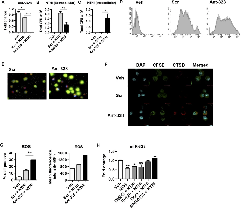
To determine whether the effects of miR-328 inhibition were due to altered phagocytosis of bacteria, or increased permissibility of phagocytes to active infection, we conducted a phagocytosis assay. Macrophages or neutrophils were exposed to heat-killed NTHi (to remove its ability to infect cells) labelled with CFSE and treated with ant-328 (or Scr control) in vitro for 1 h, and uptake assessed both by flow cytometry and fluorescence microscopy. Inhibition of miR-328 function in macrophages (Fig 2D and 2E) and neutrophils (S3D Fig) substantially enhanced phagocytosis of heat-killed NTHi (significantly increased levels of intracellular CFSE). The use of trypan blue in these experiments to quench CFSE fluorescence from extracellular bacteria attached to the cell surface confirmed the increased intracellular uptake after ant-328 treatment. To directly demonstrate that ant-328 increased the phagocytosis of bacteria we pre-treated macrophages with both antagomir and cytochalasin D (a potent phagocytosis inhibitor) before exposing the cells to heat-killed CFSE-labelled bacteria. Inhibition of phagocytsosis by cytochalasin D significantly reduced the effect of ant-328 on uptake of bacteria (S4 Fig). The marked effect on bacterial clearance by ant-328 was not related to apoptosis since there were no significant difference in the rate of cell death following antagomir treatment in either macrophages or neutrophils (S5A and S5B Fig). Treatment of macrophages with the mimetic to miR-328 had no effect on bacterial clearance likely because the basal expression level of miR-328 in lung macrophages is already high and so any additive effect becomes masked (S6A and S6B Fig). We next determined how the effects of ant-328 on enhancing phagocytosis may be regulated by examining specific bacterial binding and uptake pathways. We observed that treatment of macrophages with ant-328 significantly increased the expression of multiple bacterial uptake pathways including the LPS binding molecule CD14, the non-opsonic scavenger receptor CD36, and the bacterial adhesion integrin CD11b (S7A and S7C Fig). Collectively, this data suggests that miR-328 regulates bacterial phagocytosis in neutrophils and macrophages at least partly through the control of cell surface bacterial binding proteins.
miR-328 regulates bacterial killing pathways and its expression is controlled by p38 and JNK signalling
To mechanistically extend our observations, we looked at some important events that occurred following phagocytosis. Using confocal microscopy, we showed that pre-treatment of macrophages with ant-328 increased expression of the lysosomal enzyme, Cathepsin D (increased red fluorescence in cytoplasm), following NTHi infection compared to Scr control treated macrophages (Fig 2F). Activation of the respiratory oxidative burst and the production of ROS is another crucial event in bacterial killing by lysosomes in phagocytes. Thus, we determined whether miR-328 plays a role in activating these killing pathways in macrophages and neutrophils. Following NTHi infection, pre-treatment of macrophages with ant-328 significantly increased both the number of cells producing ROS and the intensity of its production per cell compared to Scr treated controls (Fig 2G). Likewise, the inhibition of miR-328 in neutrophils produced similar results (S3E Fig). Importantly, in neutrophils ROS production was increased by ant-328 treatment even in the absence of NTHi, although to a lower extent, suggesting that the effect on these bacterial killing pathways was partly independent of phagocytosis (S8A and S8B Fig). Interestingly, the increased killing was not associated with pro-inflammatory cell activation as the major pro-inflammatory cytokines produced by infection, IL-6 and TNF-α, were not altered by ant-328 treatment of macrophages (S9A and S9B Fig). Thus, miR-328 plays a very specific role in the microbicidal activity of these innate immune cells.
To investigate the signalling pathways involved in the regulation of miR-328 expression, we used specific inhibitors to block the activation of p38, JNK, and ERK MAPK following NTHi infection. Exposure of macrophages to NTHi or vehicle (DMSO) plus NTHi led to a significant 2-fold reduction in miR-328 expression (Fig 2H). However, pre-treatment of macrophages with a p38 inhibitor, doramapimod, or a JNK inhibitor, SP600125, prior to NTHi infection, completely blocked the down-regulation of miR-328 expression (Fig 2H). In contrast, the ERK inhibitor, U0126, had no effect. This suggests that NTHi down regulates miR-328 at least in part through p38 and JNK signalling pathways. We confirmed that NTHi exposure to primary macrophages activates p38 (S10A Fig) and JNK (S10B Fig) but not ERK (S10C Fig) signalling.
We then determined whether other miRNAs that were also identified from the array data (S1 Fig) as having increased or decreased expression in the lungs of mice following NTHi infection, could also play a role in bacterial clearance. Antagomir-mediated inhibition of miR-21-3p and miR-223 (increased expression following bacterial infection), or miR-376c (decreased expression), and miR-21 (control, no change in expression) had no effects on NTHi clearance by neutrophils in vitro (S11 Fig). Collectively, these data suggest that NTHi activates the p38 and JNK signalling pathways, which regulates the cellular levels of miR-328. Inhibition of miR-328 with ant-328 suggests that this microRNA regulates phagocytosis and killing of bacteria by macrophages and neutrophils in vitro.
MiR-328 regulates bacterial clearance by macrophages and neutrophils in vivo
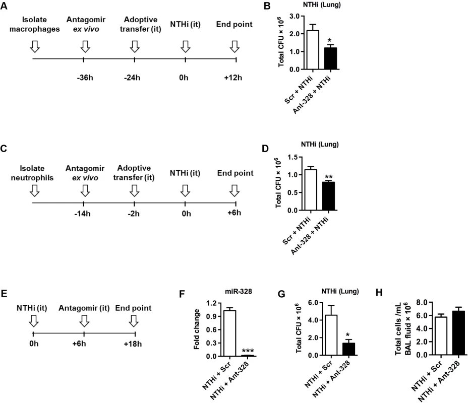
To specifically assess the role of miR-328 in macrophage- and neutrophil-mediated bacterial clearance in vivo we conducted adoptive transfer experiments. Macrophages or neutrophils were isolated from naïve mice and then treated with ant-328 or Scr control for 12 h before i.t. transfer into recipient naïve BALB/c mice (Fig 3A and 3C). To monitor the effectiveness of transfer, cells were pre-stained with CFSE. Mice were then inoculated with NTHi. Both CFSE-labelled macrophages (S12B and S12C Fig) and neutrophils (S12E and S12F Fig) entered the lungs and remained there for at least 24 and 2 h, respectively. There were no differences in the numbers of these cells between ant-328- and Scr-treated controls. Total bacterial load in BAL fluid and lung homogenates was measured by plating and colony counting. Importantly, mice that received ant-328 treated macrophages (Fig 3A and 3B) or neutrophils (Fig 3C and 3D) showed significantly improved clearance of NTHi from the lungs compared to Scr control treatment. However, there were no statistical differences in total inflammatory cell infiltrates in BAL fluid (S13A and S13F Fig). Mice that received ant-328 treated macrophages had similar total number of macrophages (S13B Fig) but a significantly reduced number of neutrophils (S13C Fig). It is likely that the reduction in neutrophil numbers is due to increased clearance of bacteria by ant-328 which leads to reduction in the early inflammatory events that promote neutrophil influx. The adoptive transfer of macrophages is likely to promote the removal of apoptotic neutrophils as well. Mice that received ant-328 treated neutrophils had no difference in macrophages (S13G Fig) or neutrophils numbers (S13H Fig). The concentrations of pro-inflammatory cytokines, IL-6 and TNF-α, in BAL fluid were equivalent in all experiments (S13D–S13E and S13I–S13J Fig). These data suggest that the effect of ant-328 on bacterial clearance was mediated directly by phagocytes and not by increased inflammatory cell recruitment or cytokine production.
Inhibition of miR-328 in the lungs promotes bacterial clearance
We next assessed whether inhibition of miR-328 improved NTHi clearance in vivo, which would indicate whether the use of ant-328 would be a potential therapeutic option for respiratory bacterial infection. Naïve BALB/c mice were inoculated with NTHi, and 6 h later were treated i.t. with ant-328 or Scr control (Fig 3E). Inhibition of miR-328 function by ant-328 was confirmed during infection (Fig 3F). NTHi clearance from the lungs was significantly enhanced (4-fold) by ant-328 treatment compared to controls (Fig 3G). Again total (Fig 3H) and neutrophils infiltration (S13L Fig) in the BAL and the levels of the pro-inflammatory cytokines, IL-6 and TNF-α, were not significantly altered (S13M–S13N Fig). In contrast, there was a small but significant increase in the number of macrophages recruited to the lungs by ant-328 treatment following NTHi infection (S13K Fig). We then tested whether the small changes in inflammatory cell numbers in the BAL in some experiments was due to the direct effect of ant-328, or indirectly due to a secondary effect induced by alterations in NTHi clearance after ant-328 treatment. In order to assess this we treated naïve mice with Scr or ant-328 and measured inflammatory cells and cytokines in the BAL. We observed that there was no effect of ant-328 on BAL cell recruitment or pro-inflammatory cytokine production in non-infected mice (S14A–S14D Fig).
Inhibition of miR-328 overcomes corticosteroid-induced immune suppression to clear bacteria
The mainstay treatment of many respiratory diseases is the prolonged use of corticosteroids, however this leads to immunosuppression and increased risk of hospitalisation for pneumonia, especially in COPD patients [23]. This has been shown in rodent and human studies to result from the suppression of phagocytosis of bacteria by macrophages [24–26].
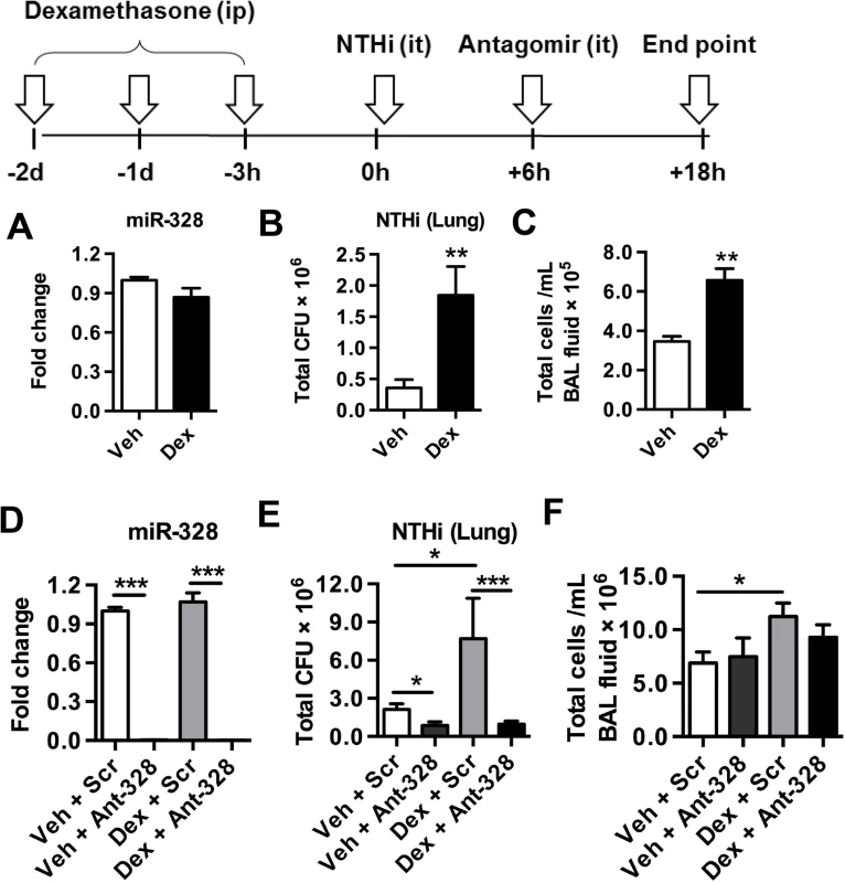
We next determined whether miR-328 was involved in dexamethasone-mediated suppression of immunity and bacterial clearance, and if inhibition of this miRNA could enhance bacterial clearance in these immune suppressed mice. Mice were administered dexamethasone i.p. daily for 3 days and were then inoculated with NTHi (scheme Fig 4). Dexamethasone treatment did not alter miR-328 expression, suggesting that this miRNA was not directly involved in mediating the effects of corticosteroids (Fig 4A). As expected, dexamethasone pre-treated mice inoculated with NTHi had substantially increased bacterial load in their lungs during infection (Fig 4B). Interestingly, pre-treatment also increased the inflammatory cell infiltrate in the lungs. This is likely due to the increased bacterial load, which results in a greater proinflammatory response leading to the recruitment of more inflammatory cells (Fig 4C). Ant-328 treatment inhibited miR-328 in both dexamethasone and vehicle-treated mice infected with NTHi to similar levels (Fig 4D). Inhibition of miR-328 in dexamethasone pre-treated mice reversed the increased bacteria load compared to controls (Fig 4E). Importantly, bacterial levels were reduced to below those in non-immune-suppressed mice (vehicle+Scr control), and to equivalent levels compared to non-immune suppressed mice treated with ant-328 (vehicle+ant-328). These data indicate that the inhibition of miR-328 was as effective at clearing bacteria in both immune competent and immune suppressed environments. Again the number of infiltrating inflammatory cells in the BAL was only increased by dexamethasone treatment and there was no difference between Scr control and the ant-328 treated groups (Fig 4F).
Inhibition of miR-328 promotes bacterial clearance in a model of cigarette smoke-induced emphysema
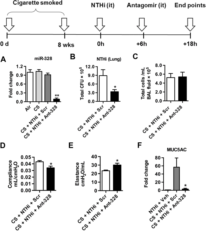
NTHi is commonly isolated from the airways of COPD patients and is linked to exacerbations of disease [20]. Thus, we next investigated if inhibition of miR-328 improved bacterial clearance in our model of cigarette smoke-induced emphysema. Mice were exposed for cigarette smoke for 8 weeks and were then inoculated with NTHi followed by antagomir treatment (scheme Fig 5). Exposure of the lung to ant-328 resulted in significant reduction in the levels of miR-328 (Fig 5A). Again, treatment with ant-328 during infection enhanced NTHi clearance in cigarette smoke-exposed mice (Fig 5B). Total numbers of cellular infiltrates were similar between ant-328 treated and Scr control groups (Fig 5C). In the same model, pulmonary function was measured using the forced oscillation technique. Inhibition of miR-328 function also resulted in a decrease in pulmonary compliance (Fig 5D) and an increase in pulmonary elastance (Fig 5E). Inhibition also nearly completely ablated the increase in muc-5ac expression induced by smoke-exposure with NTHi infection (Fig 5F).
MiR-328 regulates phagocytosis by human macrophages and neutrophils
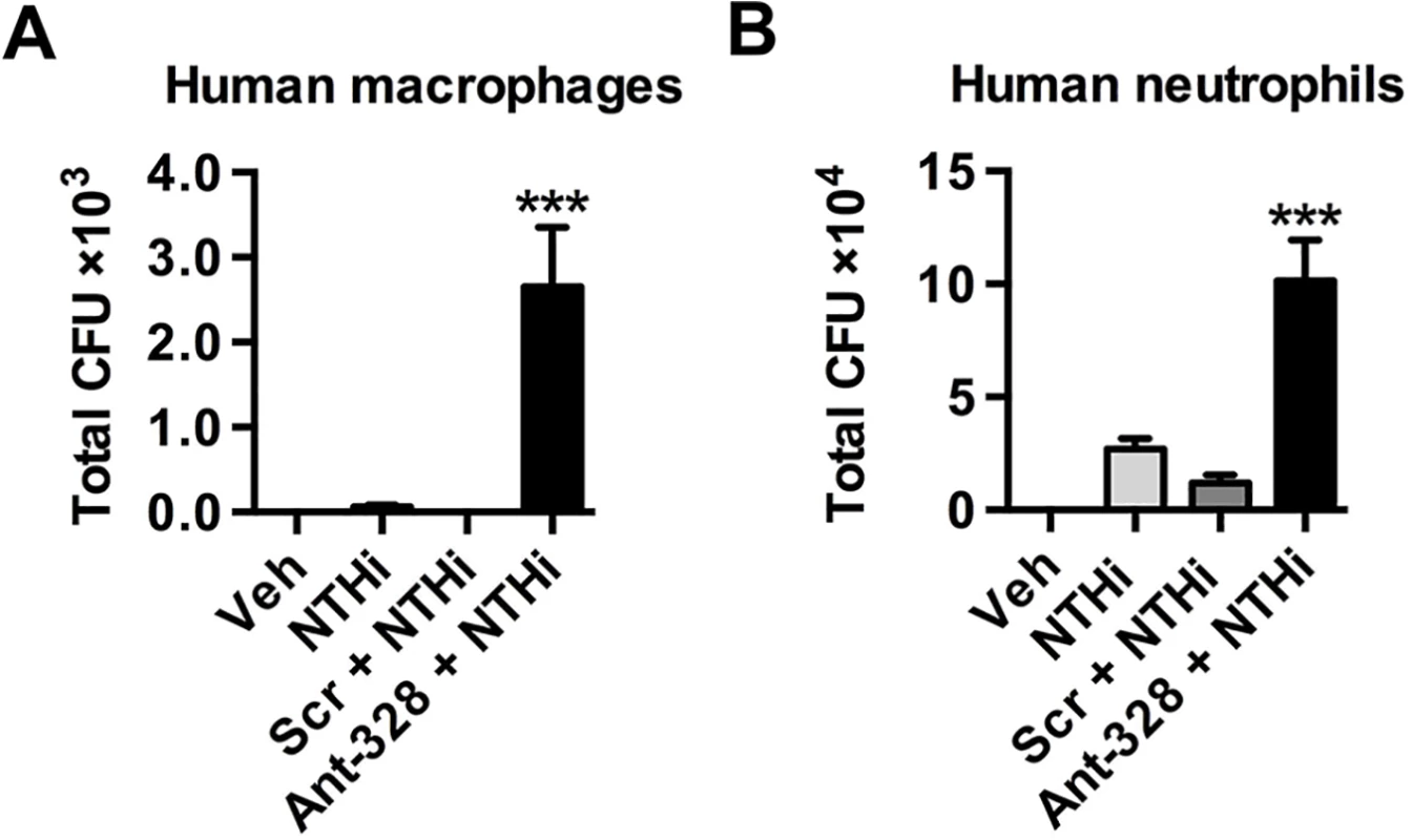
MiR-328 is conserved across species, with an identical sequence in mice and humans. Therefore, we investigated if miR-328 plays a similar role in human macrophages and neutrophils infected with NTHi in vitro. Neutrophils were purified from healthy adult blood and macrophages were differentiated in culture from monocytes derived from the PBMC fraction. Similar to the results observed with murine cells, inhibition of miR-328 significantly increased bacterial uptake by human macrophages (Fig 6A) and neutrophils (Fig 6B) in vitro.
Discussion
miRNAs are known to be involved in regulating innate immune responses. Expression is induced by various stimuli such as TLR pathways [27,28] or pro-inflammatory cytokines [29,30] and they can act as both positive and negative feedback signals to control these pathways. Furthermore, host pathogen interactions and activation of the immune system has been shown to alter, and be altered by, miRNA expression in various infection models [31–35]. In this study we show that within 24 h of NTHi infection the levels of miRNA in the lung are rapidly altered with expression of individual miRNAs being both increased and decreased. Alterations in the level of expression are strongly associated with bacterial clearance and recruitment of innate immune cells into the lung. Notably from profiling data, miR-328, a highly conserved miRNA, was significantly down-regulated by infection and its basal level are amongst the highest of all down-regulated miRNA, suggesting a key role in the host response to NTHi infection. Similarly, infection of primary lung macrophages by NTHi also downregulated miR-328 expression in vitro.
miR-328 is involved in cancer [36–38], autoimmunity [39], and neuronal disease [40]. Here we demonstrate a new role in regulating innate immune cell function, wherein suppression of miR-328 improves bacterial clearance in the lungs. Inhibition of this miRNA in either macrophages or neutrophils from both mice and humans in vitro increases uptake (of live and heat-killed NTHi) and decreases survival of bacteria. This indicates that miR-328 regulates phagocytosis and killing of bacteria. Bacterial clearance is also dependent on ROS production [41]. In chronic granulomatous disease, phagocytic activity is normal, however, there is an inability of phagocytes to form ROS after phagocytosis, which leads to increased bacterial survival [42]. Here we show that increased production of ROS in ant-328 treated phagocytes, indicating that this bacterial killing mechanism is also regulated by miR-328. Oxygen-independent bacterial killing mechanisms (such as Cathepsin D) also operate following phagocytosis although they usually coincide with oxygen-dependent lysosomal killing. Cathepsin D is a lysosomal cationic protease that disrupts bacterial membranes, and increases susceptibility of gram-negative bacteria to lysis by lysozyme [43]. We observed increased cathepsin D expression in ant-328 treated macrophages suggesting that bacterial killing was also enhanced using an oxygen independent pathway. Thus miR-328 plays a critical role in the microbial host defence mechanism of innate immune cells by augmenting phagocytosis, the production of ROS and microbicidal activity. Although inhibition with an antagomir dramatically promoted NTHi clearance, application of a mimetic (increasing miR-328 levels) failed to delay clearance of NTHi. We speculate that this is because the levels of endogenous miR-328 are very high, hence masking the effect of mimetic treatment. High levels of miR-328 are linked to macrophage homeostasis and thus it is only when levels are dramatically decreased by antagomir treatment that the cell becomes activated to clear bacteria.
In cancer, miR-328 expression is regulated through the ERK1/2 pathway [38]. Here we found that p38 and JNK MAPK is activated by NTHi infection, and inhibition of these MAPK signalling prior to bacterial infection prevents the down-regulation of miR-328 levels. This suggests that NTHi suppresses miR-328 expression via activation of the p38 and JNK MAPK pathway. We did not observe a functional role for ERK. These experiments were performed in vitro with macrophages. Thus, it is possible that miR-328 expression could be regulated by other pathways in a more complex in vivo environment.
Adoptive transfer of miR-328-depleted macrophages or neutrophils increased bacterial clearance in the lung, further supporting our in vitro observations and directly demonstrating that inhibition of miR-328 in these cells amplifies their ability to clear respiratory infections. We next explored the potential of ant-328 as a therapeutic treatment for bacterial lung infection by directly administering the inhibitor to the lung after NTHi inoculation. Inhibition of miR-328 with ant-328 substantially increased bacterial clearance suggesting that targeting this miRNA could be potentially used as a new approach to anti-microbial therapy. Although we detected increased ROS production in vitro following ant-328 treatment, which is often linked to lung injury and activation of pro-inflammatory pathways, we did not observe any increase in inflammation as measured by cellular infiltration and the production of pro-inflammatory cytokines in the lung. One potential explanation is that the increased ROS production that occurs with more rapid bacterial clearance following ant-328 treatment does not reach a threshold level or period of production that is required to elicit deleterious inflammatory changes in the lung. Thus, importantly, treatment with ant-328 in vivo enhanced bacterial clearance without a consequent negative impact on lung inflammation (cellular recruitment or proinflammatory mediator production) or pathology.
In chronic lung disease, corticosteroid treatment can lead to immunosuppression and increase the risk of pneumonia [23]. In our current study pre-treatment with dexamethasone prior to NTHi infection significantly impaired bacterial clearance from the lungs. Previous studies have shown that dexamethasone inhibits macrophage phagocytosis in vitro while treatment in vivo suppresses clearance of bacteria [24,25]. Importantly, dexamethasone administration did not alter miR-328 expression levels despite the increased bacterial load in the lungs. Notably, ant-328 treatment of dexamethasone immune-suppressed mice brought about a dramatic reduction in bacterial load, to levels below that seen in immune competent mice. This data demonstrates that although dexamethasone treatment inhibits phagocytosis along with other inflammatory anti-bacterial pathways, this effect is overcome by treatment with ant-328. These results suggest that it may be possible to use ant-328 in conjunction with dexamethasone in the treatment of chronic lung diseases in order to better control bacterial infection. This would be of particular interest in the case of antibiotic resistant bacterial strains where conventional therapies tend to fail and in patients receiving steroid therapy where infections are difficult to control.
Bacterial colonisation and infection are commonly associated with COPD and exacerbation of disease, and NTHi is one of the most frequently isolated strains of bacteria [20]. The reason underlying this may be that alveolar macrophages from COPD patients exposed to cigarette smoke extract are less efficient at phagocytosing NTHi [44,45]. Using a cigarette smoked-induced model of experimental COPD [46], we demonstrated that inhibiting miR-328 brought about a 3-fold increase in bacterial clearance. A definitive characteristic of COPD is the loss of elastic recoil in the lung and increased lower airway remodelling including mucous cell hyperplasia [47]. Following infection with NTHi, treatment with ant-328 significantly improved both elastic recoil and suppressed muc-5ac expression in the lungs of chronically cigarette exposed mice. The exact mechanism of how this occurs remains to be elucidated but it may involve altered surface tension in lungs by impairing mucus production [48]. In a clinical study on acute exacerbation of COPD, half of the sputum samples from patients enrolled tested positive for bacterial growth. Approximately 50% of the strains isolated were H. influenzae and Moraxella catarrhalis, of which the vast majority were resistant to penicillin [49]. MiR-328 has a highly conserved sequence between mice and humans. Inhibition of miR-328 function in human macrophages and neutrophils using ant-328 also resulted in increased bacterial phagocytosis, indicating that our studies are potentially translatable into anti-microbial innate host defence pathways in human cells.
Our study identifies a potential alternative approach to the treatment of pathogenic microbial infections and bacterial induced exacerbation of chronic lung disease. Although the specific targets of miR-328 remain to be elucidated it is likely that this miRNA targets transcripts involved in regulating phagocytosis and controlling the microbicidal activity of the macrophages and neutrophils. Importantly, ant-328 enhances bacterial killing by specifically augmenting bacterial clearance pathways without further promoting a proinflammatory environment that may be deleterious to lung tissue. Targeting miRNA would be of particular interest to enhance therapeutic outcomes in patients suffering from disorders such as COPD, cystic fibrosis and asthma where innate host defences may be compromised due to steroid therapy or underlying disease mechanisms. Innate immune defects also occur in HIV and transplantation patients. In HIV patients, phagocytosis and respiratory burst are reduced in monocytes and neutrophils [50]. After solid organ transplantation ~50% of deaths are due to infection and during the first four months following bone marrow transplantation, phagocytosis by alveolar macrophage phagocytosis is impaired [51,52]. Although speculative, targeting miR-328 may be more broadly applicable and lead to improvement of innate immune cell function in these immune-deficient patients. While the employment of direct inhibitors of miRNA pathways needs to be approached with caution, we would envisage acute application of antagomir to treat infection or exacerbations induced by acute infections. It will be important, however, to study the chronic impact of altering miR-328 function in murine models, and subsequently in human disease. To date we have not observed toxic effects of antagomirs in chronic models of asthma [53]. In a similar approach, treatment of chronically infected chimpanzees with a locked nucleic acid–modified oligonucleotide (SPC3649) complementary to miR-122 induced long-lasting suppression of HCV viremia, with no evidence of viral resistance or side effects in the treated animals [54]. However, continued research into the amazing regulatory powers of these small non-coding RNA is required to fully determine their potential for the treatment of disease and their fundamental role in maintaining health.
This study is the first to identify a role for miR-328 in regulating bacterial infection in the lung and provides proof-of-principle data that targeting specific host miRNAs could lead to future therapeutics to combat multi-drug resistant bacteria and infection in chronic lung diseases and immune-compromised patients.
Materials and Methods
Ethics statement
The animal protocols used were conducted in accordance with the NSW, Australia Animal Research legislation. The experimental protocols 987, A-2009-152, A-2009-141 have been reviewed and approved by University of Newcastle Animal Ethics Committee. All adult subjects provided their informed written consent to participate in the study. All human studies were conducted in accordance with approval from the University of Newcastles Human Research Ethics Committee.
Mice and NTHi
Specific pathogen-free adult male BALB/c mice were used throughout, and were obtained from the University of Newcastle central animal house. NTHi biotype II (NTHi-289) was obtained, prepared and used as previously described [55,56]. Bacteria were prepared prior to infection by growing overnight on chocolate agar plates (Oxoid, Australia) at 37°C in an atmosphere of 5% CO2. The following day, the bacteria were harvested, washed and diluted in sterile PBS and CFU calculated by spectrophotometric analysis. Live NTHi were used in NTHi infection both in vivo and in vitro. Heat killed bacteria were generated by incubating the live NTHi at 70°C for 30min.
NTHi infection in vivo
Mice were anaesthetised (12.5 mg/kg Alfaxan, Jurox, NSW, Australia) intravenously via the tail vein and innoculated intratracheally (i.t) using a cathether (Terumo, Sureflo Hospital Supplies of Australia) with 5x10⁵ or 5x10⁶ CFU of live NTHi in 30 μl of PBS. Following NTHi inoculation, mice were sacrificed at various time points and BAL fluid and lung homogenate (homogenised in 1 ml sterile PBS (Gibco, Invitrogen, New Zealand)) were used to determine bacterial growth. Serial dilutions were loaded onto chocolate agar plates (Oxoid, Australia) and incubated overnight at 37°C in an atmosphere of 5% CO2. After 16 h, bacterial colonies for NTHi were counted. BAL cells were enumerated as described previously [57].
miRNA quantitative RT-PCR
Total RNA was isolated using TRI Reagent (Ambion). miRNA expression was validated using TaqMan miRNA assays (Applied Biosystem) according to the manufacturer’s protocol and normalised to the housekeeping small RNA gene sno-202.
Measurement of respiratory burst by flow cytometry
Macrophages in cell culture were detached in citric saline (0.135 M KCL, 0.015 M Na citrate) while neutrophils were removed by rinsing. Cells were spun down and resuspended in FACS buffer (2% fetal calf serum (FCS; Sigma-Aldrich) in PBS) before incubation with 10μM dihydroethidium (DHE; Sigma-Aldrich) for 30 minutes at 37°C. Fluorescence intensity was measured by flow cytometry with 488nm excitation and 610nm emission.
Immunofluorescence staining
Immunofluorescence assays were performed as described previously [56]. Macrophages were labelled with 5μM of CFSE (Molecular Probes) for 10 minutes at 37°C. Staining was quenched with fetal calf serum (FCS) for 1 minute, and cells were washed and resuspended in PBS. Cells were fixed in 1% (w/v) paraformaldehyde in PBS buffer for 10 min at room temperature (RT), blocked and permeabilized with 0.2% (v/v) Triton X-100 and 5% normal goat serum for 1 h at RT. Cathepsin D (CTSD) expression were stained using rabbit monoclonal Ab to CTSD (Abcam) overnight at 4°C. Cells were then incubated with Cy3-conjugated goat anti-rabbit IgG (GE Healthcare) for 45 minutes at RT and nucleus was counter-stained with DAPI (Sigma-Aldrich). Images were analyzed using Olympus FluoView FV1000 confocal microscope.
Inhibition of p38 MAPK signalling pathways
Macrophages were pre-treated 5μM doramapimod (inhibit p38 MAPK) (LC lab), 50μM SP600125 (inhibit JNK) (LC lab), 50μM U0126 (inhibit ERK) (LC lab) for 30 minutes,prior to NTHi infection for 8 h. DMSO was used as a vehicle control.
Cigarette smoke exposure
Mice were exposed to cigarette smoke (in the form of 12 cigarettes) in a closed chamber twice a day, 5 days per week for a duration of 8 weeks as previously described [46].
Dexamethasone treatment
Mice were injected intraperitoneally (i.p.) with 3mg/kg of dexamethasone (DEX; Sigma-Aldrich) for 3 consecutive days before they were inoculated with (5 x 10⁵ CFU / mouse) with NTHi.
Antagomir oligonucleotides
The complementary antagomir strand of the miR-328-3p sequence obtained from miRbase was synthesised from Sigma-Aldrich with the following chemical modifications 5’mA.*.mC.*.mG.mG.mA.mA.mG.mG.mG.mC.mA.mG.mA.mG.mA.mG.mG.mG.mC.*.mC.*.mA.*.mG.*. 3’-Chol where “m” represents 2’-OMe-modified phosphoramidites, “*” represents phosphorothioate linkages, and “-Chol” was hydroxyprolinol-linked cholesterol to allow permeation of cell membranes. The scrambled control antagomir sequence was 5'mU.*.mC.*.mA.mC.mA.mA.mC.mC.mU.mC.mC.mU.mA.mG.mA.mA.mA.mG.mA.*.mG.*.mU.*.mA.*.3'-Chol. Mice were treated in vivo with 50μg of antagomir intratracheally (see NTHi infection: methods), and macrophages and neutrophils were treated with antagomir at 50μg/ml 12 h before assays in vitro.
Statistical analysis
Results are presented as means ± standard errors of the means (SEM). Results were analysed using one way ANOVA plus Bonferroni post-test for statistical significance or Student's t-test plus Mann-Whitney test. Analysis was performed with Prism v4.0 (GraphPad Software, CA. USA). p< 0.05 was considered to represent a significant difference.
Additional methods were described in S1 Methods.
Supporting Information
Zdroje
1. Mizgerd JP (2006) Lung infection—a public health priority. PLoS Med 3: e76. 16401173
2. Barnes PJ, Drazen JM, Rennard SI, Thomson NC (2008) Asthma and COPD Basic Mechanisms and Clinical Management. 2nd ed. San Diego: Academic Press Imprint Elsevier Science & Technology Books.
3. Monso E, Ruiz J, Rosell A, Manterola J, Fiz J, et al. (1995) Bacterial infection in chronic obstructive pulmonary disease. A study of stable and exacerbated outpatients using the protected specimen brush. Am J Respir Crit Care Med 152: 1316–1320. 7551388
4. Simpson JL, Grissell TV, Douwes J, Scott RJ, Boyle MJ, et al. (2007) Innate immune activation in neutrophilic asthma and bronchiectasis. Thorax 62: 211–218. 16844729
5. Thimmulappa RK, Gang X, Kim JH, Sussan TE, Witztum JL, et al. (2012) Oxidized phospholipids impair pulmonary antibacterial defenses: evidence in mice exposed to cigarette smoke. Biochem Biophys Res Commun 426: 253–259. doi: 10.1016/j.bbrc.2012.08.076 22935414
6. Talbot TR, Hartert TV, Mitchel E, Halasa NB, Arbogast PG, et al. (2005) Asthma as a risk factor for invasive pneumococcal disease. N Engl J Med 352: 2082–2090. 15901861
7. Rinehart JJ, Sagone AL, Balcerzak SP, Ackerman GA, LoBuglio AF (1975) Effects of corticosteroid therapy on human monocyte function. N Engl J Med 292: 236–241. 1089191
8. Rennie RP, Ibrahim KH (2005) Antimicrobial resistance in Haemophilus influenzae: how can we prevent the inevitable? Commentary on antimicrobial resistance in H. influenzae based on data from the TARGETed surveillance program. Clin Infect Dis 41 Suppl 4: S234–238. 16032558
9. Hoban DJ, Doern GV, Fluit AC, Roussel-Delvallez M, Jones RN (2001) Worldwide prevalence of antimicrobial resistance in Streptococcus pneumoniae, Haemophilus influenzae, and Moraxella catarrhalis in the SENTRY Antimicrobial Surveillance Program, 1997–1999. Clin Infect Dis 32 Suppl 2: S81–93. 11320449
10. Baltimore D, Boldin MP, O'Connell RM, Rao DS, Taganov KD (2008) MicroRNAs: new regulators of immune cell development and function. Nat Immunol 9: 839–845. doi: 10.1038/ni.f.209 18645592
11. Foster PS, Plank M, Collison A, Tay HL, Kaiko GE, et al. (2013) The emerging role of microRNAs in regulating immune and inflammatory responses in the lung. Immunol Rev 253: 198–215. doi: 10.1111/imr.12058 23550648
12. Bartel DP (2004) MicroRNAs: genomics, biogenesis, mechanism, and function. Cell 116: 281–297. 14744438
13. Bazzoni F, Rossato M, Fabbri M, Gaudiosi D, Mirolo M, et al. (2009) Induction and regulatory function of miR-9 in human monocytes and neutrophils exposed to proinflammatory signals. Proc Natl Acad Sci U S A 106: 5282–5287. doi: 10.1073/pnas.0810909106 19289835
14. Taganov KD, Boldin MP, Chang KJ, Baltimore D (2006) NF-kappaB-dependent induction of microRNA miR-146, an inhibitor targeted to signaling proteins of innate immune responses. Proc Natl Acad Sci U S A 103: 12481–12486. 16885212
15. Domingo-Gonzalez R, Katz S, Serezani CH, Moore TA, Levine AM, et al. (2013) Prostaglandin E2-induced changes in alveolar macrophage scavenger receptor profiles differentially alter phagocytosis of Pseudomonas aeruginosa and Staphylococcus aureus post-bone marrow transplant. J Immunol 190: 5809–5817. doi: 10.4049/jimmunol.1203274 23630358
16. Wang J, Yang K, Zhou L, Minhaowu, Wu Y, et al. (2013) MicroRNA-155 promotes autophagy to eliminate intracellular mycobacteria by targeting Rheb. PLoS Pathog 9: e1003697. doi: 10.1371/journal.ppat.1003697 24130493
17. Liu PT, Wheelwright M, Teles R, Komisopoulou E, Edfeldt K, et al. (2012) MicroRNA-21 targets the vitamin D-dependent antimicrobial pathway in leprosy. Nat Med 18: 267–273. doi: 10.1038/nm.2584 22286305
18. Ma F, Xu S, Liu X, Zhang Q, Xu X, et al. (2011) The microRNA miR-29 controls innate and adaptive immune responses to intracellular bacterial infection by targeting interferon-gamma. Nat Immunol 12: 861–869. doi: 10.1038/ni.2073 21785411
19. Murphy TF, Sethi S (1992) Bacterial infection in chronic obstructive pulmonary disease. Am Rev Respir Dis 146: 1067–1083. 1416398
20. Monso E, Ruiz J, Rosell A, Manterola J, Fiz J, et al. (1995) Bacterial infection in chronic obstructive pulmonary disease. A study of stable and exacerbated outpatients using the protected specimen brush. American journal of respiratory and critical care medicine 152: 1316–1320. 7551388
21. Nelson S, Summer WR (1998) Innate immunity, cytokines, and pulmonary host defense. Infect Dis Clin North Am 12: 555–567, vii. 9779378
22. Reynolds HY (1983) Lung inflammation: role of endogenous chemotactic factors in attracting polymorphonuclear granulocytes. Am Rev Respir Dis 127: S16–25. 6221680
23. Ernst P, Gonzalez AV, Brassard P, Suissa S (2007) Inhaled Corticosteroid Use in Chronic Obstructive Pulmonary Disease and the Risk of Hospitalization for Pneumonia. American Journal of Respiratory and Critical Care Medicine 176: 162–166. 17400730
24. Nakamura Y, Murai T, Ogawa Y (1996) Effect of in vitro and in vivo administration of dexamethasone on rat macrophage functions: comparison between alveolar and peritoneal macrophages. European Respiratory Journal 9: 301–306. 8777968
25. Becker J, Grasso RJ (1985) Suppression of phagocytosis by dexamethasone in macrophage cultures: inability of arachidonic acid, indomethacin, and nordihydroguaiaretic acid to reverse the inhibitory response mediated by a steroid-inducible factor. Int J Immunopharmacol 7: 839–847. 3935587
26. Herzer P, Lemmel EM (1980) Inhibition of granulocyte function by prednisolone and non-steroid anti-inflammatory drugs. Quantitative evaluation with NBT test and its correlation with phagocytosis. Immunobiology 157: 78–88. 6894133
27. Taganov KD, Boldin MP, Chang KJ, Baltimore D (2006) NF-kappaB-dependent induction of microRNA miR-146, an inhibitor targeted to signaling proteins of innate immune responses. Proceedings of the National Academy of Sciences of the United States of America 103: 12481–12486. 16885212
28. Liu G, Friggeri A, Yang Y, Park YJ, Tsuruta Y, et al. (2009) miR-147, a microRNA that is induced upon Toll-like receptor stimulation, regulates murine macrophage inflammatory responses. Proceedings of the National Academy of Sciences of the United States of America 106: 15819–15824. doi: 10.1073/pnas.0901216106 19721002
29. Tili E, Michaille J-J, Cimino A, Costinean S, Dumitru CD, et al. (2007) Modulation of miR-155 and miR-125b Levels following Lipopolysaccharide/TNF-α Stimulation and Their Possible Roles in Regulating the Response to Endotoxin Shock. The Journal of Immunology 179: 5082–5089. 17911593
30. O'Connell RM, Taganov KD, Boldin MP, Cheng G, Baltimore D (2007) MicroRNA-155 is induced during the macrophage inflammatory response. Proceedings of the National Academy of Sciences 104: 1604–1609. 17242365
31. Liu PT, Wheelwright M, Teles R, Komisopoulou E, Edfeldt K, et al. (2012) MicroRNA-21 targets the vitamin D-dependent antimicrobial pathway in leprosy. Nature medicine 18: 267–273. doi: 10.1038/nm.2584 22286305
32. Schulte LN, Eulalio A, Mollenkopf HJ, Reinhardt R, Vogel J (2011) Analysis of the host microRNA response to Salmonella uncovers the control of major cytokines by the let-7 family. The EMBO journal 30: 1977–1989. doi: 10.1038/emboj.2011.94 21468030
33. Xiao B, Liu Z, Li BS, Tang B, Li W, et al. (2009) Induction of microRNA-155 during Helicobacter pylori infection and its negative regulatory role in the inflammatory response. The Journal of infectious diseases 200: 916–925. doi: 10.1086/605443 19650740
34. Izar B, Mannala GK, Mraheil MA, Chakraborty T, Hain T (2012) microRNA Response to Listeria monocytogenes Infection in Epithelial Cells. International journal of molecular sciences 13: 1173–1185. doi: 10.3390/ijms13011173 22312311
35. Cremer TJ, Ravneberg DH, Clay CD, Piper-Hunter MG, Marsh CB, et al. (2009) MiR-155 induction by F. novicida but not the virulent F. tularensis results in SHIP down-regulation and enhanced pro-inflammatory cytokine response. PLoS One 4: e8508. doi: 10.1371/journal.pone.0008508 20041145
36. Arora S, Ranade AR, Tran NL, Nasser S, Sridhar S, et al. (2011) MicroRNA-328 is associated with (non-small) cell lung cancer (NSCLC) brain metastasis and mediates NSCLC migration. International journal of cancer Journal international du cancer 129: 2621–2631. doi: 10.1002/ijc.25939 21448905
37. Pan YZ, Morris ME, Yu AM (2009) MicroRNA-328 negatively regulates the expression of breast cancer resistance protein (BCRP/ABCG2) in human cancer cells. Molecular pharmacology 75: 1374–1379. doi: 10.1124/mol.108.054163 19270061
38. Eiring AM, Harb JG, Neviani P, Garton C, Oaks JJ, et al. (2010) miR-328 functions as an RNA decoy to modulate hnRNP E2 regulation of mRNA translation in leukemic blasts. Cell 140: 652–665. doi: 10.1016/j.cell.2010.01.007 20211135
39. Padgett KA, Lan RY, Leung PC, Lleo A, Dawson K, et al. (2009) Primary biliary cirrhosis is associated with altered hepatic microRNA expression. Journal of autoimmunity 32: 246–253. doi: 10.1016/j.jaut.2009.02.022 19345069
40. Boissonneault V, Plante I, Rivest S, Provost P (2009) MicroRNA-298 and microRNA-328 regulate expression of mouse beta-amyloid precursor protein-converting enzyme 1. The Journal of biological chemistry 284: 1971–1981. doi: 10.1074/jbc.M807530200 18986979
41. Rada BK, Geiszt M, Kaldi K, Timar C, Ligeti E (2004) Dual role of phagocytic NADPH oxidase in bacterial killing. Blood 104: 2947–2953. 15251984
42. Quie PG, White JG, Holmes B, Good RA (1967) In vitro bactericidal capacity of human polymorphonuclear leukocytes: diminished activity in chronic granulomatous disease of childhood. The Journal of clinical investigation 46: 668–679. 6021213
43. Thorne KJ, Oliver RC, Barrett AJ (1976) Lysis and killing of bacteria by lysosomal proteinases. Infect Immun 14: 555–563. 971964
44. Berenson CS, Garlipp MA, Grove LJ, Maloney J, Sethi S (2006) Impaired Phagocytosis of Nontypeable Haemophilus influenzae by Human Alveolar Macrophages in Chronic Obstructive Pulmonary Disease. Journal of Infectious Diseases 194: 1375–1384. 17054066
45. Martí-Lliteras P, Regueiro V, Morey P, Hood DW, Saus C, et al. (2009) Nontypeable Haemophilus influenzae Clearance by Alveolar Macrophages Is Impaired by Exposure to Cigarette Smoke. Infection and Immunity 77: 4232–4242. doi: 10.1128/IAI.00305-09 19620348
46. Beckett EL, Stevens RL, Jarnicki AG, Kim RY, Hanish I, et al. (2013) A new short-term mouse model of chronic obstructive pulmonary disease identifies a role for mast cell tryptase in pathogenesis. J Allergy Clin Immunol 131: 752–762. doi: 10.1016/j.jaci.2012.11.053 23380220
47. BALDI S, MINIATI M, BELLINA CR, BATTOLLA L, CATAPANO G, et al. (2001) Relationship between Extent of Pulmonary Emphysema by High-resolution Computed Tomography and Lung Elastic Recoil in Patients with Chronic Obstructive Pulmonary Disease. American Journal of Respiratory and Critical Care Medicine 164: 585–589. 11520720
48. Ingenito EP, Tsai LW, Majumdar A, Suki B (2005) On the role of surface tension in the pathophysiology of emphysema. Am J Respir Crit Care Med 171: 300–304. 15563634
49. Larsen MV, Janner JH, Nielsen SD, Friis-Moller A, Ringbaek T, et al. (2009) Bacteriology in acute exacerbation of chronic obstructive pulmonary disease in patients admitted to hospital. Scandinavian journal of infectious diseases 41: 26–32. doi: 10.1080/00365540802484828 18855228
50. Michailidis C, Giannopoulos G, Vigklis V, Armenis K, Tsakris A, et al. (2012) Impaired phagocytosis among patients infected by the human immunodeficiency virus: implication for a role of highly active anti-retroviral therapy. Clinical and experimental immunology 167: 499–504. doi: 10.1111/j.1365-2249.2011.04526.x 22288593
51. Corensek MJ, Stewart RW, Keys TF, Mehta AC, McHenry MC, et al. (1988) A multivariate analysis of risk factors for pneumonia following cardiac transplantation. Transplantation 46: 860–865. 3061082
52. Winston DJ, Territo MC, Ho WG, Miller MJ, Gale RP, et al. (1982) Alveolar macrophage dysfunction in human bone marrow transplant recipients. The American journal of medicine 73: 859–866. 6756138
53. Collison A, Herbert C, Siegle JS, Mattes J, Foster PS, et al. (2011) Altered expression of microRNA in the airway wall in chronic asthma: miR-126 as a potential therapeutic target. BMC Pulm Med 11: 29. doi: 10.1186/1471-2466-11-29 21605405
54. Lanford RE, Hildebrandt-Eriksen ES, Petri A, Persson R, Lindow M, et al. (2010) Therapeutic silencing of microRNA-122 in primates with chronic hepatitis C virus infection. Science 327: 198–201. doi: 10.1126/science.1178178 19965718
55. Essilfie AT, Simpson JL, Horvat JC, Preston JA, Dunkley ML, et al. (2011) Haemophilus influenzae infection drives IL-17-mediated neutrophilic allergic airways disease. PLoS Pathog 7: e1002244. doi: 10.1371/journal.ppat.1002244 21998577
56. Essilfie AT, Simpson JL, Dunkley ML, Morgan LC, Oliver BG, et al. (2012) Combined Haemophilus influenzae respiratory infection and allergic airways disease drives chronic infection and features of neutrophilic asthma. Thorax 67: 588–599. doi: 10.1136/thoraxjnl-2011-200160 22387445
57. Foster PS, Hogan SP, Ramsay AJ, Matthaei KI, Young IG (1996) Interleukin 5 deficiency abolishes eosinophilia, airways hyperreactivity, and lung damage in a mouse asthma model. J Exp Med 183: 195–201. 8551223
Štítky
Hygiena a epidemiologie Infekční lékařství LaboratořČlánek vyšel v časopise
PLOS Pathogens
2015 Číslo 4
- Jak souvisí postcovidový syndrom s poškozením mozku?
- Měli bychom postcovidový syndrom léčit antidepresivy?
- Farmakovigilanční studie perorálních antivirotik indikovaných v léčbě COVID-19
- 10 bodů k očkování proti COVID-19: stanovisko České společnosti alergologie a klinické imunologie ČLS JEP
Nejčtenější v tomto čísle
- Toxin-Induced Necroptosis Is a Major Mechanism of Lung Damage
- Transgenic Fatal Familial Insomnia Mice Indicate Prion Infectivity-Independent Mechanisms of Pathogenesis and Phenotypic Expression of Disease
- Role of Hypoxia Inducible Factor-1α (HIF-1α) in Innate Defense against Uropathogenic Infection
- EphrinA2 Receptor (EphA2) Is an Invasion and Intracellular Signaling Receptor for
Zvyšte si kvalifikaci online z pohodlí domova
Současné možnosti léčby obezity
nový kurzVšechny kurzy
