-
Články
Reklama
- Vzdělávání
- Časopisy
Top články
Nové číslo
- Témata
Reklama- Videa
- Podcasty
Nové podcasty
Reklama- Kariéra
Doporučené pozice
Reklama- Praxe
ReklamaLocal and Systemic Regulation of Plant Root System Architecture and Symbiotic Nodulation by a Receptor-Like Kinase
Despite the essential functions of roots in plant access to water and nutrients, root system architecture has not been directly considered for crop breeding improvement, but it is now considered key for a “second green revolution.” In this study, we aimed to decipher integrated molecular mechanisms coordinating lateral organ development in legume roots: lateral roots and nitrogen-fixing symbiotic nodules. The compact root architecture 2 (cra2) mutant form an increased number of lateral roots and a reduced number of symbiotic nitrogen-fixing nodules. This mutant is affected in a CLAVATA1-like Leucine-Rich Repeat Receptor-Like Kinase (LRR-RLK) that has not previously been linked to root development. Grafting experiments showed that CRA2 negatively controls lateral root formation and positively controls nodule development through local and systemic pathways, respectively. Overall, our results can be integrated in the framework of regulatory pathways controlling the symbiotic nodule number, the so-called “Autoregulation of Nodulation” (AON), involving another LRR-RLK that also acts systemically from the shoots, SUNN (Super Numeric Nodules). A coordinated function of the CRA2 and SUNN LRR-RLKs may thereby permit the dynamic fine tuning of the nodule number depending on the environmental conditions.
Published in the journal: . PLoS Genet 10(12): e32767. doi:10.1371/journal.pgen.1004891
Category: Research Article
doi: https://doi.org/10.1371/journal.pgen.1004891Summary
Despite the essential functions of roots in plant access to water and nutrients, root system architecture has not been directly considered for crop breeding improvement, but it is now considered key for a “second green revolution.” In this study, we aimed to decipher integrated molecular mechanisms coordinating lateral organ development in legume roots: lateral roots and nitrogen-fixing symbiotic nodules. The compact root architecture 2 (cra2) mutant form an increased number of lateral roots and a reduced number of symbiotic nitrogen-fixing nodules. This mutant is affected in a CLAVATA1-like Leucine-Rich Repeat Receptor-Like Kinase (LRR-RLK) that has not previously been linked to root development. Grafting experiments showed that CRA2 negatively controls lateral root formation and positively controls nodule development through local and systemic pathways, respectively. Overall, our results can be integrated in the framework of regulatory pathways controlling the symbiotic nodule number, the so-called “Autoregulation of Nodulation” (AON), involving another LRR-RLK that also acts systemically from the shoots, SUNN (Super Numeric Nodules). A coordinated function of the CRA2 and SUNN LRR-RLKs may thereby permit the dynamic fine tuning of the nodule number depending on the environmental conditions.
Introduction
Plant growth requires the continuous development of the root system and its adaptation to changing environmental soil conditions. Mechanisms controlling root system architecture at the whole-plant level, including the systemic coordination of shoot and root development, are key breeding targets for maintaining crop productivity under adverse stress conditions but remain poorly understood [1]. Root system architecture is a consequence of the sustained activity of root apical meristems, leading to indeterminate root growth as well as the de novo formation of lateral organs. In legume (Fabaceae) plants, the root system can form two types of lateral organs depending on the environmental conditions: lateral roots and symbiotic nitrogen-fixing nodules [2]–[4]. Lateral root initiation, emergence and growth depend on water and nutrient availability and are regulated by a combination of local and systemic pathways [5]. Symbiotic nodules are formed under nitrogen-deprived conditions when the specific Rhizobium spp. soil bacteria are present in the rhizosphere [6]–[7]. In both types of lateral organogeneses, cell divisions are activated in specific tissues (pericycle, endodermis and cortex) above the growing root tip [2]–[4], [8]. Root tissues contributing to primordium formation are, however, different depending on the plants and organs: in the Medicago truncatula model legume, lateral root primordia mainly develop from pericycle cell divisions, whereas the nodule primordia that are induced by Sinorhizobium meliloti mainly derive from the inner cortex. Both types of primordia will then subsequently emerge from the parental root and establish a meristematic stem cell niche ensuring their indeterminate growth.
To control meristematic activity, cell differentiation, and lateral organ initiation, non-cell autonomous cues are essential to carry positional information, which can be informed either by mobile phytohormones, small RNAs or peptides [9]–[11]. Among peptides, several CLAVATA 3/EMBRYO-SURROUNDING REGION (like) peptides (so called CLE peptides; [10]) that are perceived by Leucine-Rich Repeats – Receptor-Like Kinases (LRR-RLKs) are involved in local and long-distance (systemic) pathways controlling the development of different plant organs. First, several CLE peptide/LRR-RLK receptor modules carry positional information across a few cell layers to control the cell fate in different Arabidopsis thaliana meristems. The founding example is the CLAVATA3 (CLV3) peptide, which is perceived by the CLV1 receptor to control the shoot apical meristem stem cell niche [12], [13] as well as columella cell differentiation in the Root Apical Meristem (RAM; [14]–[16]). A second example is the Tracheary element Differentiation Inhibitory Factor (TDIF) peptide, which is perceived by the Phloem Intercalated with Xylem (PXY) receptor, controlling stem cell proliferation/differentiation transition in the cambium meristem and therefore vasculature differentiation and organ thickening [17]–[19].
In legume plants, an additional CLE/LRR-RLK module controlling root lateral organs number was identified through grafting experiments as performing a long-distance systemic function from the shoots [7]. Mutants affecting orthologous LRR-RLKs that are closely related to CLV1 in Arabidopsis (SUNN, Super Numeric Nodules in M. truncatula; NARK, Nodule Autoregulation receptor Kinase in soybean; and HAR1, Hypernodulation and Aberrant Root 1 in Lotus japonicus; [20]–[23]) form an increased number of symbiotic nitrogen-fixing nodules depending on the receptor function in the shoots. In addition, the Lotus KLAVIER (KLV) LRR-RLK, which is closely related to the Arabidopsis RECEPTOR-LIKE PROTEIN KINASE 2 (RPK2)/TOADSTOOL 2 (TOAD2) that is functionally linked to anther and embryo development, is involved in the same systemic AON pathway as is HAR1 [24]. In M. truncatula and L. japonicus, CLE peptides that are specifically produced in nodulated roots can negatively regulate the nodule number depending on these LRR-RLK receptors [25]–[27]. This CLE peptide/LRR-RLK module therefore participates in the systemic “Autoregulation of Nodulation” (AON; [22]) pathway, which may involve a direct root-to-shoot CLE peptide transport and receptor binding in the shoots, as recently proposed [28]. Interestingly, an increased number of emerged lateral roots was reported in the Lotus har1 mutant under both symbiotic and non-symbiotic conditions [29]. Similarly, KLV also has a non-symbiotic function in the local regulation of SAM maintenance [24]. Collectively, these results indicate that CLE peptide/LRR-RLK signaling modules regulate the development of various organs using either local and/or systemic pathways.
Results
The COMPACT ROOT ARCHITECTURE 2 (CRA2) gene negatively regulates lateral root development
Forward genetic screens that were performed on a M. truncatula Tnt1 insertional mutant collection [30]–[31] identified seven mutant lines with a wild-type shoot development and a “compact root system architecture” (so called “cra”; Fig. 1A–C and S1A Fig.). Segregation analyses of this root phenotype revealed a 3∶1 WT:mutant ratio (chi2 test, p<0.05, n = 30), suggestive of a single locus recessive mutation. Allelism tests (S1B Fig.) indicated that all of the mutants were affected in the same locus but differed from the previously identified cra1 mutant showing partially similar phenotypes [32] and were therefore named cra2. Detailed quantitative in vitro analyses revealed that the cra2 phenotype consists, compared to the WT, of shorter roots with an increased number of emerged lateral roots (Fig. 1D–E). Accordingly, lateral roots were observed three days post germination (dpg; Fig. 1E) in contrast to WT plants. This root phenotype was observed independently of the growth and nutrient conditions that were used (greenhouse versus in vitro, Fig. 1A, E; with or without nitrogen or carbon sources, Fig. 1E). In addition to the faster emergence of lateral roots, we observed a reduction in the primary root growth, which prompted us to analyze the structure of the RAM (Fig. 2A–C). In contrast to the A. thaliana model, M. truncatula roots have an open meristem, and the transition between cell proliferation and differentiation is more progressive. In addition, cells from diverse files elongate at a slightly different distance from the root apex, leading to a cone-shaped transition zone (Fig. 2A, detail of the transition zone in S2 Fig.). When cra2 RAMs were observed three dpg, both the cell proliferation and elongation zones were reduced, and a lower number of cells was observed in the two zones (Fig. 2 A–C). Root patterning, however, evaluated based on both longitudinal and transversal sections, seemed unaffected (Fig. 2A–B and D). In addition, amyloplast accumulation in differentiated root cap cells and the expression of the RAM stem cell niche marker WOX5 (WUSCHEL-related homeobox 5; [33]–[35]) were both detected in cra2 (S3 Fig.), suggesting the maintenance of RAM cell identity. Among several hypotheses that could explain the cra2 root system architecture phenotype, we tested whether a RAM activity defect could indirectly lead to increased branching as compensation or if the reduced meristematic activity could be a consequence of the enhanced formation of lateral roots. An analysis of root apices at one dpg, i.e., before any lateral root primordium could be detected in the cra2 mutant, revealed no significant difference in the size of the cell proliferation and elongation zones between WT and mutant roots (Fig. 2E, F). Compared to previous observations of roots at three dpg (Fig. 2A, B), this result indicates that a RAM defect does not precede the occurrence of the lateral root phenotype. As an independent approach, we experimentally removed the RAM at one or three dpg and followed the kinetics of lateral root formation (Fig. 2G, H). The cra2 mutant showed an increased ability to form lateral roots whether RAM excision occurred before or after lateral root initiation. Collectively, these results suggest that the cra2 RAM phenotype can be disconnected from its enhanced ability to form lateral roots.
Fig. 1. The “compact root architecture 2” (cra2) mutants have short roots and more lateral roots independently of the growth conditions. 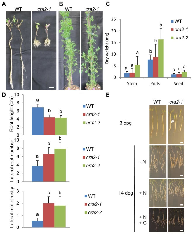
A and B, Representative examples of wild-type (WT) and cra2-1 plants that were grown in the greenhouse for one month on a perlite-sand mixture (A) or for three months on soil (B). Bar = 1 cm in A, 10 cm in B. C, Quantification of the stems, pods and seeds dry weight of the WT, cra2-1 and cra2-2 plants that are shown in (B). The error bars represent confidence intervals (α = 5%). A Kruskal and Wallis test was used to determine the significant differences (indicated by letters, α<5%; n = 10). D, Quantification of the root length (upper graph), lateral root number (middle graph) and lateral root density (lower graph) of the WT, cra2-1 and cra2-2 plants that were grown in vitro for 10 days post-germination (dpg) on an N-deprived “i” medium [42]. The error bars represent confidence intervals (α = 1%). A Kruskal and Wallis test was used to determine the significant differences (indicated by letters, α<1%; n>25). E, Representative examples of the WT and cra2-1 plants that were grown in vitro for three dpg on an N-deprived “i” medium [42] or for 14 dpg on the same medium (- N), on an N-rich medium (+N, Fahraeus with NH4NO3 10 mM; [43]), or on an N- and C-rich medium (+N+C, “Lateral Root Inducing Medium; [42]). Note that in cra2, the lateral roots were already emerged at three dpg (arrowhead) and that the “compact root system architecture” phenotype was detectable independently of the growth medium. Bars = 0,5 cm. Fig. 2. The cra2 lateral root and root apical meristem phenotypes can be disconnected. 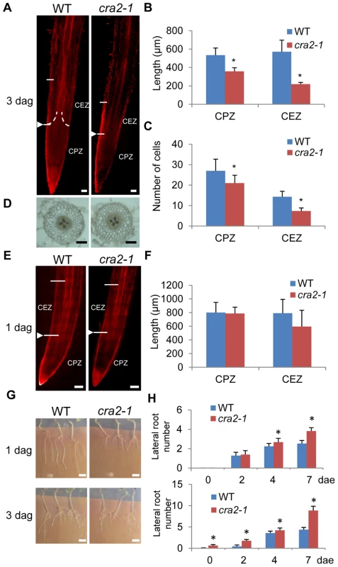
A and E, Representative examples of the wild-type (WT) and cra2-1 Root Apical Meristems (RAM, stained with Propidium Iodide to visualize cell walls) three days post germination (dpg; A) or one dpg (E). The arrowhead indicates the apical position of the “cone-shaped” transition zone (as defined in S2 Fig.) between the cell proliferation zone (CPZ) and the cell elongation zone (CEZ). Bar = 100 µm. B, F, Quantification in the WT and cra2-1 roots of CPZ and CEZ length at three dpg (B) or one dpg (F). C, Quantification in the WT and cra2-1 roots of the cells number in the same zones at three dpg. In B, C, and F, the error bars represent standard deviation, and a Mann-Whitney test was used to determine the significant differences between genotypes (*, α<5%, n = 10). D, Transversal sections of three dpg WT and cra2-1 roots showing a similar radial organization of cell layers. Bar = 100 µm. G, Representative examples of the WT and cra2-1 root system architecture seven days post excision (dpe) of the RAM. The excision was performed either one day after germination (dpg, upper pictures) or three dpg (lower pictures). Bar = 1 cm. H, Quantification of the lateral roots at two, four and seven days post excision (dpe) of the RAM in WT or cra2-1 plants. As shown in (G), the meristems were excised at either one dpg (upper graph) or three dpg (lower graph). The error bars represent the confidence interval (α = 5%), and a Mann-Whitney test was used to determine significant differences between the genotypes for each time point (*, α<5%; n>25). The CRA2 gene positively regulates symbiotic nodule development
In addition to root branching, legume roots can adapt to environmental conditions by developing another root lateral organ, the nitrogen-fixing symbiotic nodule. An analysis of cra2 roots under symbiotic conditions revealed that a similar “compact root architecture” phenotype was observed (Fig. 3A) and that mutant shoot growth was similar with or without Rhizobium inoculation (Fig. 3B, C). cra2 plants, however, developed a strongly reduced number of symbiotic nodules (Fig. 3 D, E). This low nodulation phenotype could be either linked to a direct CRA2 function in regulating nodulation or may reflect that the strongly altered “compact root architecture” phenotype (e.g., Fig. 3A) indirectly hampers nodule formation. Using a symbiotic infection kinetic analysis (1 to 14 dpg), we showed that Rhizobium inoculation as early as 1 or 3 dpg led to reduced nodulation (Fig. 3E), indicating that the cra2 nodulation phenotype was independent of the strength of the lateral root phenotype. The few symbiotic nodules that formed on cra2 roots were elongated (S4A Fig.), revealing that a functional nodule meristem was formed. In addition, an analysis of bacterial nitrogenase activity using an Acetylene Reduction Assay (ARA) showed that despite cra2 plants have a strongly reduced ability to fix atmospheric nitrogen due to their lower number of symbiotic organs (S4B Fig.), cra2 nodules have a similar specific ARA activity compared to that of WT nodules (S4C Fig.). Overall, the detailed analysis of the mutant phenotypes indicates that, independently of a potential indirect effect of the increased lateral root formation on nodule formation, CRA2 antagonistically regulates lateral root and nodule formation.
Fig. 3. The cra2 mutants have reduced symbiotic nodulation. 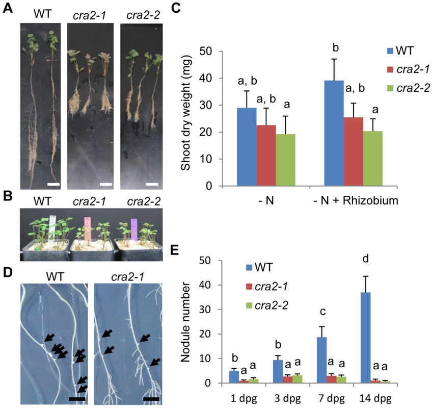
A, Representative examples of the wild-type (WT), cra2-1 and cra2-2 plants that were grown in the greenhouse on a perlite-sand mixture for two weeks (A) or one month (B) on an N-deprived medium (“i”, [42]) and then inoculated by Sinorhizobium meliloti 1021 for 14 days. Bar = 1 cm. C, Quantification of the shoot dry weights of the WT, cra2-1 and cra2-2 plants that are shown in (A). The error bars represent confidence intervals (α = 5%). A Kruskal and Wallis test was used to determine the significant differences (indicated by letters, α<5%; n = 30). D, Representative examples of the WT and cra2-1 plants that were grown for one week in vitro on an N-deprived medium (“i”, [42]) and then inoculated by S. meliloti 1021 for 14 days. The arrowheads indicate the position of the nodules. Bar = 1 cm. E, Quantification of the nodule number 14 days post inoculation with S. meliloti in the roots of the WT and cra2-1 plants that were grown in vitro. The inoculation was performed between one and 14 days post germination (dpg). The error bars represent confidence intervals (α = 5%). Significant differences were determined using a Mann-Whitney test (α<5%; n>20). CRA2 regulates root system architecture depending on systemic and local pathways
As root system architecture is controlled by both systemic and local pathways, we next determined using graftings experiments whether the cra2 phenotypes depend on roots and/or shoots. Under non-symbiotic conditions, the “compact root architecture” phenotype was recovered specifically when the graft combination included cra2 mutant roots (Fig. 4A–B) but was independent of the shoot genotype, indicating that CRA2 expression in roots is sufficient to negatively regulate lateral root formation. Under symbiotic conditions, we surprisingly observed a disconnect between the lateral root and nodulation phenotypes (Fig. 4C–D). Indeed, similar to non-symbiotic conditions, the increased density of the lateral roots was associated with cra2 mutant roots, but the low nodulation phenotype was observed only in grafting combinations that included cra2 mutant shoots and was independent of the root genotype. This result indicates that the systemic activity of CRA2 in the shoots positively regulates symbiotic nodule formation in the roots. Strikingly, the cra2/WT grafts had a WT root system architecture but developed more than 10 times fewer nodules (Fig. 4C–D). In addition, when the nodule numbers were related to the root dry weight to counterbalance the strong differences existing between WT/WT and WT/cra2 root systems, the nodulation efficiency became strictly equivalent between these two grafting combinations (Fig. 4E). These results therefore unambiguously demonstrate that the low-nodulation phenotype is not an indirect consequence of an inhibitory signal that is produced in the numerous lateral roots in cra2. In addition, this result indicates that the root-dependent regulation of the lateral root number is independent of the shoot-regulation of the nodule number.
Fig. 4. CRA2 locally regulates lateral root formation and systemically regulates symbiotic nodule formation from the shoots. 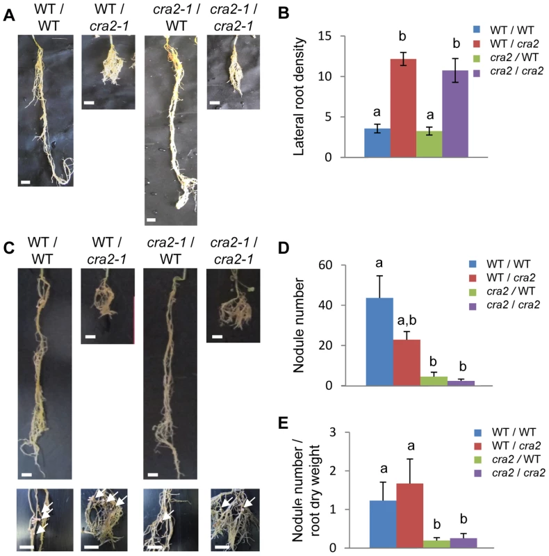
A and C, Representative images of different grafting combinations between the wild-type (WT) and cra2-1 plants that were grown in greenhouse with a perlite-sand mixture for eight weeks on an N-rich medium (A) or on an N-deprived medium with Sinorhizobium meliloti 1021 (C). The shoot/root grafting combinations are indicated above each picture. In panel (C), detailed pictures showing nodules (arrows) are included below. Bar = 1 cm. B, Quantification of the lateral root density (number of emerged lateral roots/centimeter of parental root) in the different grafting combinations that are shown in (A). D and E, Quantification of the nodule numbers (D) and the nodule number related to the root dry weight (E) in the different grafting combinations that are shown in (C). In B, D and E, the error bars represent confidence intervals (α = 5%), and the significant differences were determined using a Kruskal and Wallis test (indicated by letters, α<5%; n>10). The CRA2 gene encodes a Leucine-Rich Repeat – Receptor-Like Kinase that has not previously been linked to root or symbiotic nodule development
To identify the gene that is affected in cra2 mutants, Tnt1 Flanking Sequence Tags (FSTs) were generated in the different alleles (cra2-1 to cra2-7) to detect Tnt1 insertions affecting a common genomic sequence in several of these mutant lines (cra2-1, cra2-4 to cra2-6; Fig. 5A and S5A–B Fig.). Interestingly, the three other alleles also showed genetic lesions in the same locus, consisting, respectively, in the presence of other M. truncatula endogenous insertional elements for cra2-3 and cra2-7 and in a point mutation for cra2-2 leading to a frameshift (S5C-E Fig.). Based on the FSTs that were available from the Noble foundation (http://bioinfo4.noble.org/mutant/), we could identify three additional lines with a Tnt1 insertion in the same open reading frame (cra2-8 to cra2-10) that showed a “compact root architecture” phenotype (Fig. 5A and S1A Fig.), further confirming that mutation at this locus is causal for the phenotype. The genomic region that was affected in the 10 available cra2 alleles encodes an LRR-RLK belonging to subfamily XI (Fig. 5B), which, in agreement with the results of the grafting experiment, is expressed in the shoots, roots and nodules (S6 Fig.). Interestingly, this LRR-RLK subfamily contains several other receptors that were identified as regulating plant development via local or systemic regulation [10], [36]. An analysis of CRA2 homology with other LRR-RLKs that have been functionally characterized in legumes showed that CRA2 was not closely related to SUNN/HAR1 or KLV and was most closely related to A. thaliana XIP1 (Xylem Intermixed in Phloem 1; [37]; Fig. 5B). An Arabidopsis xip1 mutant was recently described as showing defects in vasculature patterning specifically in the shoots but not in the roots, and no root developmental phenotype was reported [37]. To determine whether XIP1 and CRA2 may nevertheless have related functions, we analyzed the vascular patterning in cra2 mutant roots and shoots. No significant defect could be detected in the shoot or root cambium structure; in the vascular bundle differentiation, such as the misspecification of xylem and phloem bundles; or in the root or stele diameter (S7 Fig.). These results suggest that XIP1 and CRA2 are not functional homologs. In addition, these results indicate that the cra2 root and nodulation phenotypes (Fig. 1, 2) are not linked to any detectable defect in vascular bundle patterning. We then analyzed the CRA2 spatial expression pattern under non-symbiotic and symbiotic conditions using either a transcriptional fusion between an ∼2 kb CRA2 promoter region and the GUS reporter (Fig. 6A–D and I–N) or in situ hybridization (Fig. 6 E–H). Both approaches revealed an expression that was associated with the root stele and vascular bundles (Fig. 6A–B and E–F). Similarly, CRA2 expression was associated with vascular bundles in the shoots (Fig. 6G–H). This result agrees with the expression pattern of other LRR-RLKs regulating lateral root and nodule numbers in different legumes (HAR1/NARK/SUNN or KLV; 24, [38]–[40]). CRA2 was additionally expressed in the Cell Proliferation Zone (CPZ) of the open RAM (Fig. 6A and C–D). In addition, CRA2 was expressed in the lateral root initiation site as soon as the pericycle divisions could be identified (Fig. 6I–J) and later in whole lateral root primordia during the initiation and emergence stages (Fig. 6K–L). Under symbiotic nodulation conditions, CRA2 expression was also detected in nodule primordia (Fig. 6M) as well as in mature nodules in relation to peripheral vascular bundles and the apical meristem (Fig. 6N).
Fig. 5. The CRA2 gene encodes a Leucine-Rich Repeat Receptor-Like Kinase (LRR-RLK). 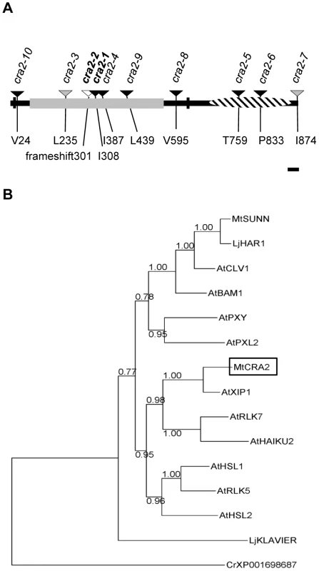
A, Structure of the CRA2 protein indicating the 10 mutant alleles (arrowheads) that were identified by forward and reverse genetic screens (the indicated position is related to the predicted ATG) and functional domains. The vertical black bars indicate the predicted transmembrane domains; in grey are the Leucine-Rich Repeats; and the hatched region represents the kinase domain. The black arrowheads represent alleles that are linked to a Tnt1 retro-element insertion; the grey arrowheads represent another insertional element; and the white arrowhead represents a nucleotide deletion causing a translational frameshift. Bar = 50 residues. B, Phylogenetic tree of selected LRR-RLKs that are related to CRA2 from Arabidopsis (subfamily XI) or are functionally characterized in legumes. The sequences were aligned using Muscle, and the regions that were conserved between all of the sequences were defined with Gblocks. The phylogenetic relationships were determined using a maximum likelihood analysis (PhyML), and statistical support for each node was estimated by approximate likelihood ratio tests. The Chlamydomonas reinhardtii XP001698687 protein was used to root the tree. Fig. 6. CRA2 expression in the shoot, root and symbiotic nodules. 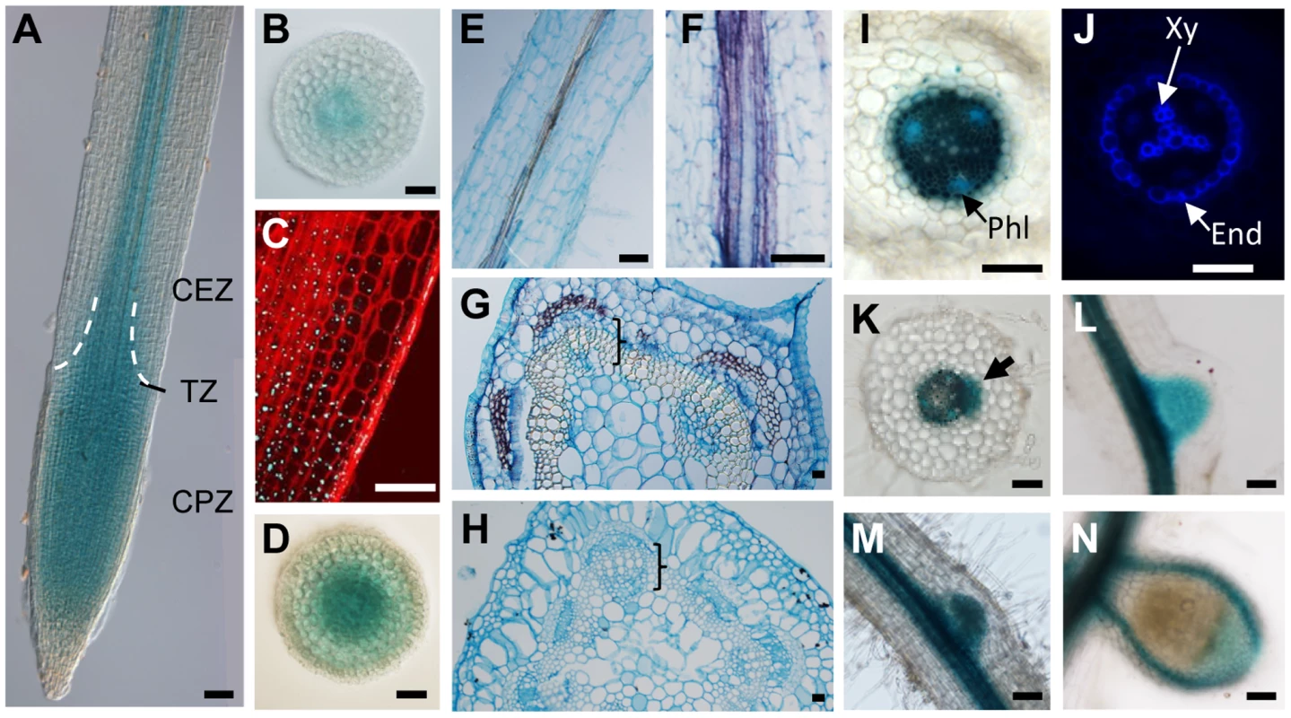
The spatial expression pattern of CRA2 was analyzed using a promoter (1,8 kb)-GUS fusion (A–D and I–N) or by in situ hybridization (E–H). A, A root apex observed in bright field with dichroic illumination (Nomarski). The dotted lines indicate the position of the “cone-shaped” transition zone (TZ) between the cell proliferation zone (CPZ) and the cell elongation zone (CEZ). B and D, Root transversal sections in the CEZ (B) and the CPZ (D). C, Detail of the root meristem transition zone using a z-stack projection in confocal sections. The cell walls are visualized with a Propidium Iodide counterstaining, and GUS staining appears in reflectance as blue dots. E–F, In situ hybridization of the CRA2 transcripts in root longitudinal sections. (F) Detail of the purple signal associated with vasculature strands. G–H, In situ hybridization of the CRA2 transcripts in stem transversal sections (G, purple signal) or with a sense probe used as a negative control (H). Brackets indicate vascular bundles. I–J, Detail of the stele in the root differentiated region in bright field (I) to visualize the GUS staining (in dark blue) and phloem vascular bundles poles (Phl, in turquoise blue) or under UV illumination (J, same section as I) to visualize the blue autofluorescence of the xylem vascular bundle poles (Xy) and endodermis (End). K–L, Lateral root primordium initiation (K, arrow) and emergence (L) observed in bright field. K is a root transversal section. M–N, Nodule primordium (M, three days post-inoculation [dpi] with S. meliloti 1021) and mature nitrogen-fixing nodule (N, 14 dpi) observed in bright field. Bars = 100 µm. Discussion
CRA2 functions in the regulation of legume root system architecture suggest that this LRR-RLK acts positively in the Autoregulation of Nodulation (AON) pathway. In the current model, the systemic SUNN-dependent AON pathway represses the number of nodules that are formed on the roots depending on the shoot metabolic status, e.g., the amount of carbon skeletons that are provided through photosynthesis and that are required for the subsequent assimilation of fixed nitrogen in the nodules. The SUNN pathway therefore limits the formation of extra nodules depending on environmental cues. When initiated, however, a negative feedback mechanism would be necessary to reset this negative regulation and further permit new symbiotic infection events. The CRA2 LRR-RLK may then participate in the systemic dynamic fine tuning of nodule formation. As both CRA2 and SUNN LRR-RLKs are expressed in the shoot vasculature, it remains to be determined whether and how these proteins act together or independently. As LRR-RLKs have been proposed to form large complexes comprising different members of the family [36] or even other RLKs (e.g., CLV1 with ACR4 in the RAM; [16]), CRA2 and SUNN may interact within a single complex.
The Lotus HAR1-dependent AON pathway was additionally shown to control negatively lateral root formation [20]–[29]. In the sunn Medicago mutant, however, the root length is reduced, but no specific function in the regulation of lateral root number has been identified [23]. CRA2 therefore negatively affects lateral roots similarly to HAR1 in Lotus but dissimilarly to SUNN in M. truncatula. Interestingly, a shorter root length and a decreased number of lateral roots occurred in the Lotus klv mutant, whereas additive effects of klv and har1 were identified for nodulation, indicating their related function in a single AON pathway [40]. In addition, both har1 and klv antagonistic lateral root phenotypes were observed under non-symbiotic conditions [29], [40], similar to the phenotype of CRA2 in M. truncatula. This result indicates that these peptide/LRR-RLK regulatory modules acting under non-symbiotic conditions may also control lateral root formation in non-nodulating (non-legume) plants. In Arabidopsis, the ACR4 CRINKLY-RLK regulates lateral root initiation [41], but no LRR-RLK has been yet associated with this developmental process. More generally, despite the close relationships between HAR1, SUNN and CLV1, their mutant phenotypes are not conserved between plant species: the sunn and har1 mutants have divergent lateral root phenotypes, whereas no shoot fasciation phenotype was detected in the har1 or sunn mutant in contrast to clv1 in Arabidopsis. Similarly, despite CRA2 and XIP1 having closely related sequences, no vasculature patterning phenotype was identified in cra2 compared to xip1, and no altered root system architecture phenotype was reported in xip1. This result suggests that various sets of LRR-RLKs differentially regulate the ability of root systems to form lateral roots depending on species, even inside the legume family. Alternatively and non-exclusively, different patterns of LRR-RLK gene duplication may have occurred in the different plant genomes, generating functionally divergent or redundant pathways. These scenarios may explain the apparent phenotypic diversification that is observed.
Overall, this study demonstrates that a single LRR-RLK acts locally in roots and systemically in shoots to control root lateral organ development, thereby coordinating at the whole-plant level the plastic development of the root system depending on the changing environmental conditions. To elucidate the opposite effects of the CRA2 pathway on lateral root and nodule formation, the identification of downstream targets will be essential. Among other candidate pathways, cytokinins were previously reported to antagonistically control lateral root and symbiotic nodule formation [42]. Data on crosstalk between signaling peptides and hormones is just emerging, mainly with auxin and cytokinins [10]. Therefore, a remaining challenge will be to integrate the different peptide/receptor modules that are known to regulate lateral root and nodule formation, including the CRA2 pathway, into the framework of hormonal regulation.
Materials and Methods
Plant and bacteria material
The Medicago truncatula (Gaertn.) plants used in this study were of the R108-4 genotype. The Tnt1 insertional mutants were generated and screened at the Noble Foundation (USA; lines named NFxxx; [30]) or produced at the “Institut des Sciences du Végétal” (ISV, CNRS, Gif sur Yvette, France) and screened at the “Agroécologie” institute (INRA, Dijon, France; lines with other names than NFxxx). The seeds were scarified on sandpaper, sterilized for 20 min in bleach (12% [v/v] sodium hypochlorite), and thoroughly rinsed in sterile water. The seeds were then stratified at 4°C for one day and then germinated at 25°C in the dark on inverted water agar plates. The seedlings were grown in vitro in a growth chamber at 25°C with a 16 h light period and a 150 µE intensity on N-deprived media (“i”, [42]; or Fahraeus, [43]), on an N-rich medium (Fahraeus with NH4NO3 10 mM), or on a N - and C-rich medium (“Lateral-Root-Inducing Medium”, LRIM; [42]) depending on the experiment. Alternatively, the plants were grown in a greenhouse (25°C, 16 h 150 µE light period, 60%–70% humidity) in soil or in pots containing a perlite∶sand mixture (3∶1) and watered every two days with “i” or “SN/2” media [42], depending on the experiment. The nodulation experiments used the Sinorhizobium meliloti 1021 strain (OD600nm = 0.05) as described in [42].
Transposon display, genotyping, RT-PCR and cloning
Plant genomic DNA was extracted from the leaves as described by [44], and Flanking Sequence Tags (FSTs) that were linked to Tnt1 insertions were identified using the transposon display PCR method as described in [45] using the EcoR1 or Ase1 restriction enzymes. The transposon display, genotyping PCRs, and the sequencing of the different mutant alleles were performed using the primers that are listed in S1 Table. The RT-PCRs and real-time RT-PCRs were performed as described in [42] with the primers that are indicated in S1 Table.
To generate the MtCRA2 reporter transcriptional fusion, an 1800-bp sequence upstream of the CRA2 start codon was identified, corresponding to the upstream region of the Medtr3g110840.1 open reading frame (M. truncatula genome Mt4.0v1, http://www.jcvi.org/medicago/index.php). This region was amplified by PCR using a high-fidelity polymerase (Phusion, Thermo Scientific) and primers pCRA2-F and pCRA2-R (S1 Table). The promoter was cloned using Gateway technology initially into the pTOPO-Entry vector (Invitrogen) and then into the pkGWFS7 vector (https://gateway.psb.ugent.be/search) carrying a Green Fluorescent Protein (GFP) - β Glucuronidase (GUS) fusion downstream of the cloning recombination site.
Grafting, root apex excision, and composite plants
The graftings were performed as described in the “cuttings and grafts” chapter of the Medicago handbook (http://www.noble.org/medicagohandbook/). The grafts, which were initially generated in vitro, were transferred after three weeks to pots containing a perlite∶sand mixture (3∶1) that was imbibed with the “i” solution in a growth chamber (same conditions as described above). After three weeks, the root length and root dry weight (60°C for 48 h) were measured.
For root apex excision, the roots were sectioned at one centimeter from the root tips one or three days post germination and placed in the “i” medium in a growth chamber (same conditions as described above). The number of lateral roots was quantified one to seven days post root tip excision using the ImageJ software (http://imagej.nih.gov/ij/).
To generate composite plants, constructs were introduced into the Agrobacterium rhizogenes ARqua1 strain and used for M. truncatula root transformation as described in [46]. Transgenic roots were selected on kanamycin (25 mg/L) for two weeks, and composite plants were then transferred onto growth papers (Mega International, http://www.mega-international.com/) on “i” medium for four to six days and, depending on the experiment, were inoculated by S. meliloti as described in [42].
Microscopy analyses, histological stainings and in situ hybridization
The roots or stems were cut into 5-mm segments and immediately embedded in 3% agarose (Euromedex). A vibratome (VT1200S, Leica) was used to generate 100 µm-thick cross-sections. Toluidine blue and aniline blue (Sigma) stainings were performed by incubating sections for 3 to 5 min in 1% toluidine blue or in 0.005% aniline blue and subsequently washing the sections twice with distilled water. The sections were viewed under bright-field illumination or under UV excitation (365 nm), respectively, for toluidine blue or for aniline blue stainings using a Leica DMI 6000B inverted microscope. The phloroglucinol–HCl reagent was prepared by mixing 2 volumes of 2% (w/v) phloroglucinol (Sigma) in 95% ethanol with one volume of concentrated HCl and observation under bright-field illumination (DMI 6000B, Leica). Callose fluorescence was visualized using a 405 nm excitation, and emission was collected between 480 and 515 nm (DMI 6000B, Leica). The amyloplasts were detected using Lugol (Sigma) staining and visualization under bright field illumination.
To examine the detailed cellular organization of M. truncatula root apices, the roots were stained using a modified Pseudo Schiff-Propidium Iodide (PS-PI) staining protocol as described by [47]. Longitudinal optical sections were obtained using a Leica TCS SP2 confocal laser-scanning microscope using a 488 nm excitation, and emission was collected between 520 and 720 nm. The root meristem size was estimated as the number of cells in the outer cortex from the location of the tip to the first elongating cell using the ImageJ software. The length of the elongated cortical cells in the mature root region was also measured.
The GUS activity was revealed as described by (48] and observed under bright-field illumination (DMI 6000B, Leica). In addition, using the AOBS reflection mode of a Leica TCS SP8 spectral confocal laser-scanning microscope, the GUS precipitate reflectance was analyzed as described in [47] with a 488 nm excitation and a collection of the reflection signal between 485 and 491 nm on PS-PI counterstained samples (detected as described above).
In situ hybridization was performed as described in [48] with the probe as indicated in S1 Table.
Nitrogen fixation activity
To measure nitrogenase activity of symbiotic nodules, an Acetylene Reduction Assay (ARA) was performed on individual plants with a protocol that was derived from [49]. One month after inoculation with Rhizobium, the plants were placed into 10-ml glass vials that were sealed with rubber septa. Acetylene was injected into each vial, and after 1 h of incubation at room temperature, the produced ethylene was measured using Gas Chromatography (7820A, Agilent technologies).
Phylogenetic and statistical analyses
Phylogenetic analyses were performed using the SeaView package (v.4.4.0; [50]). The full-length protein sequences that were retrieved from the NCBI database were aligned using Muscle and optimized with the Gblocks software. Phylogenetic relationships were defined using a maximum likelihood approach. The tree was built with the PhyML software using the LG substitution model and four substitution rate categories. Support for each node was gained by approximate likelihood ratio tests (aLRT SH-like; [51]). The XP_001698687.1 RLK from Chlamydomonas patens, which was identified by a BLAST search of NCBI (E-value = 1e-40), was used as an outgroup to anchor the tree.
The statistical analyses were performed with non-parametric tests (Mann-Whitney when n = 2 independent samples and Kruskal and Wallis when n>2 independent samples).
Supporting Information
Zdroje
1. ComasLH, BeckerSR, CruzVM, ByrnePF, DierigDA (2013) Root traits contributing to plant productivity under drought. Front Plant Sci 4 : 442.
2. Gonzalez-Rizzo S, Laporte P, Crespi M, Frugier F (2009) Legume root architecture: a peculiar root system. In: Beeckman T editor, Blackwell, Oxford, United Kingdom. Annual Plant Reviews: Chapter 10, Root Development. pp.239–87.
3. DesbrossesGJ, StougaardJ (2011) Root nodulation: a paradigm for how plant-microbe symbiosis influences host developmental pathways. Cell Host Microbe 10 : 348–58.
4. OldroydGE, MurrayJD, PoolePS, DownieJA (2011) The rules of engagement in the legume-rhizobial symbiosis. Annu Rev Genet 45 : 119–44.
5. MalamyJE (2005) Intrinsic and environmental response pathways that regulate root system architecture. Plant Cell Environ 28 : 67–77.
6. CrespiM, FrugierF (2008) De novo organ formation from differentiated cells: root nodule organogenesis. Sci Signal 1: re11.
7. ReidDE, FergusonBJ, HayashiS, LinYH, GresshoffPM (2011) Molecular mechanisms controlling legume autoregulation of nodulation. Ann Bot 108 : 789–95.
8. HerrbachV, RemblièreC, GoughC, BensmihenS (2014) Lateral root formation and patterning in Medicago truncatula. J Plant Physiol 171 : 301–10.
9. FukakiH, TasakaM (2009) Hormone interactions during lateral root formation. Plant Mol Biol 69 : 437–49.
10. MurphyE, SmithS, De SmetI (2012) Small signaling peptides in Arabidopsis development: how cells communicate over a short distance. Plant Cell 24 : 3198–217.
11. ChenX (2012) Small RNAs in development - insights from plants. Curr Opin Genet Dev 22 : 361–7.
12. ClarkSE, WilliamsRW, MeyerowitzEM (1997) The CLAVATA1 gene encodes a putative receptor kinase that controls shoot and floral meristem size in Arabidopsis. Cell 89 : 575–85.
13. FletcherJC, BrandU, RunningMP, SimonR, MeyerowitzEM (1999) Signaling of cell fate decisions by CLAVATA3 in Arabidopsis shoot meristems. Science 283 : 1911–4.
14. Casamitjana-MartínezE, HofhuisHF, XuJ, LiuCM, HeidstraR, et al. (2003) Root-specific CLE19 overexpression and the sol1/2 suppressors implicate a CLV-like pathway in the control of Arabidopsis root meristem maintenance. Curr Biol 13 : 1435–41.
15. FiersM, GolemiecE, XuJ, van der GeestL, HeidstraR, et al. (2005) The 14-amino acid CLV3, CLE19, and CLE40 peptides trigger consumption of the root meristem in Arabidopsis through a CLAVATA2-dependent pathway. Plant Cell 17 : 2542–53.
16. StahlY, GrabowskiS, BleckmannA, KühnemuthR, Weidtkamp-PetersS, et al. (2013) Moderation of Arabidopsis root stemness by CLAVATA1 and ARABIDOPSIS CRINKLY4 receptor kinase complexes. Curr Biol 23 : 362–71.
17. ItoY, NakanomyoI, MotoseH, IwamotoK, SawaS, et al. (2006) Dodeca-CLE peptides as suppressors of plant stem cell differentiation. Science 313 : 842–5.
18. FisherK, TurnerS (2007) PXY, a receptor-like kinase essential for maintaining polarity during plant vascular-tissue development. Curr Biol 17 : 1061–6.
19. HirakawaY, ShinoharaH, KondoY, InoueA, NakanomyoI, et al. (2008) Non-cell-autonomous control of vascular stem cell fate by a CLE peptide/receptor system. Proc Natl Acad Sci U S A 105 : 15208–13.
20. KrusellL, MadsenLH, SatoS, AubertG, GenuaA, et al. (2002) Shoot control of root development and nodulation is mediated by a receptor-like kinase. Nature 420 : 422–6.
21. NishimuraR, HayashiM, WuGJ, KouchiH, Imaizumi-AnrakuH, et al. (2002) HAR1 mediates systemic regulation of symbiotic organ development. Nature 420 : 426–9.
22. SearleIR, MenAE, LaniyaTS, BuzasDM, Iturbe-OrmaetxeI, et al. (2003) Long-distance signaling in nodulation directed by a CLAVATA1-like receptor kinase. Science 299 : 109–12.
23. SchnabelE, JournetEP, de Carvalho-NiebelF, DucG, FrugoliJ (2005) The Medicago truncatula SUNN gene encodes a CLV1-like leucine-rich repeat receptor kinase that regulates nodule number and root length. Plant Mol Biol 58 : 809–22.
24. MiyazawaH, Oka-KiraE, SatoN, TakahashiH, WuGJ, et al. (2010) The receptor-like kinase KLAVIER mediates systemic regulation of nodulation and non-symbiotic shoot development in Lotus japonicus. Development 137 : 4317–25.
25. OkamotoS, OhnishiE, SatoS, TakahashiH, NakazonoM, et al. (2009) Nod factor/nitrate-induced CLE genes that drive HAR1-mediated systemic regulation of nodulation. Plant Cell Physiol 50 : 67–77.
26. SaurIM, OakesM, DjordjevicMA, IminN (2011) Crosstalk between the nodulation signaling pathway and the autoregulation of nodulation in Medicago truncatula. New Phytol 190 : 865–74.
27. MortierV, De WeverE, VuylstekeM, HolstersM, GoormachtigS (2012) Nodule numbers are governed by interaction between CLE peptides and cytokinin signaling. Plant J 70 : 367–76.
28. OkamotoS, ShinoharaH, MoriT, MatsubayashiY, KawaguchiM (2013) Root-derived CLE glycopeptides control nodulation by direct binding to HAR1 receptor kinase. Nat Commun 4 : 2191.
29. WopereisJ, PajueloE, DazzoFB, JiangQ, GresshoffPM, et al. (2000) Short root mutant of Lotus japonicus with a dramatically altered symbiotic phenotype. Plant J 23 : 97–114.
30. TadegeM, WenJ, HeJ, TuH, KwakY, et al. (2008) Large-scale insertional mutagenesis using the Tnt1 retrotransposon in the model legume Medicago truncatula. Plant J 54 : 335–47.
31. TadegeM, WangTL, WenJ, RatetP, MysoreKS (2009) Mutagenesis and beyond! Tools for understanding legume biology. Plant Physiol 151 : 978–84.
32. LaffontC, BlanchetS, LapierreC, BrocardL, RatetP, et al. (2010) The compact root architecture1 gene regulates lignification, flavonoid production, and polar auxin transport in Medicago truncatula. Plant Physiol 153 : 1597–607.
33. KamiyaN, NagasakiH, MorikamiA, SatoY, MatsuokaM (2003) Isolation and characterization of a rice WUSCHEL-type homeobox gene that is specifically expressed in the central cells of a quiescent center in the root apical meristem. Plant J 35 : 429–41.
34. HaeckerA, Gross-HardtR, GeigesB, SarkarA, BreuningerH, et al. (2004) Expression dynamics of WOX genes mark cell fate decisions during early embryonic patterning in Arabidopsis thaliana. Development 131 : 657–68.
35. OsipovaMA, MortierV, DemchenkoKN, TsyganovVE, TikhonovichIA, et al. (2012) Wuschel-related homeobox5 gene expression and interaction of CLE peptides with components of the systemic control add two pieces to the puzzle of autoregulation of nodulation. Plant Physiol 158 : 1329–41.
36. BetsuyakuS, SawaS, YamadaM (2011) The Function of the CLE Peptides in Plant Development and Plant-Microbe Interactions. Arabidopsis Book 9: e0149.
37. BryanAC, ObaidiA, WierzbaM, TaxFE (2012) XYLEM INTERMIXED WITH PHLOEM1, a leucine-rich repeat receptor-like kinase required for stem growth and vascular development in Arabidopsis thaliana. Planta 235 : 111–22.
38. NontachaiyapoomS, ScottPT, MenAE, KinkemaM, SchenkPM, et al. (2007) Promoters of orthologous Glycine max and Lotus japonicus nodulation autoregulation genes interchangeably drive phloem-specific expression in transgenic plants. Mol Plant Microbe Interact 20 : 769–80.
39. SchnabelE, KarveA, KassawT, MukherjeeA, ZhouX, et al. (2012) The M. truncatula SUNN gene is expressed in vascular tissue, similarly to RDN1, consistent with the role of these nodulation regulation genes in long distance signaling. Plant Signal Behav 7 : 4–6.
40. Oka-KiraE, TatenoK, MiuraK, HagaT, HayashiM, et al. (2005) klavier (klv), a novel hypernodulation mutant of Lotus japonicus affected in vascular tissue organization and floral induction. Plant J 44 : 505–15.
41. De SmetI, VassilevaV, De RybelB, LevesqueMP, GrunewaldW, et al. (2008) Receptor-like kinase ACR4 restricts formative cell divisions in the Arabidopsis root. Science 322 : 594–7.
42. Gonzalez-RizzoS, CrespiM, FrugierF (2006) The Medicago truncatula CRE1 cytokinin receptor regulates lateral root development and early symbiotic interaction with Sinorhizobium meliloti. Plant Cell 18 : 2680–93.
43. TruchetG, DebelléF, VasseJ, TerzaghiB, GarneroneAM, et al. (1985) Identification of a Rhizobium meliloti pSym2011 region controlling the host specificity of root hair curling and nodulation. J Bacteriol 164 : 1200–10.
44. d'ErfurthI, CossonV, EschstruthA, LucasH, KondorosiA, et al. (2003) Efficient transposition of the Tnt1 tobacco retrotransposon in the model legume Medicago truncatula. Plant J 34 : 95–106.
45. Ratet P, Wen J, Cosson V, Tadege M, Mysore KS (2009) Tnt1 induced mutations in Medicago: characterisation and applications. In: Meksem K and Kahl G editors, Wiley, Oxford, United Kingdom. The Handbook of Plant Mutation Screening. pp. 83–99.
46. Boisson-DernierA, ChabaudM, GarciaF, BécardG, RosenbergC, et al. (2001) Agrobacterium rhizogenes-transformed roots of Medicago truncatula for the study of nitrogen-fixing and endomycorrhizal symbiotic associations. Mol Plant Microbe Interact 14 : 695–700.
47. TruernitE, BaubyH, DubreucqB, GrandjeanO, RunionsJ, et al. (2008) High-resolution whole-mount imaging of three-dimensional tissue organization and gene expression enables the study of Phloem development and structure in Arabidopsis. Plant Cell 20 : 1494–503.
48. PletJ, WassonA, ArielF, Le SignorC, BakerD, et al. (2011) MtCRE1-dependent cytokinin signaling integrates bacterial and plant cues to coordinate symbiotic nodule organogenesis in Medicago truncatula. Plant J 65 : 622–33.
49. KochB, EvansHJ (1966) Reduction of acetylene to ethylene by soybean root nodules. Plant Physiol 41 : 748–50.
50. GouyM, GuindonS, GascuelO (2010) SeaView version 4: A multiplatform graphical user interface for sequence alignment and phylogenetic tree building. Mol Biol Evol 27 : 221–4.
51. GuindonS, DufayardJF, LefortV, AnisimovaM, HordijkW, et al. (2010) New algorithms and methods to estimate maximum-likelihood phylogenies: assessing the performance of PhyML 3.0. Syst Biol 59 : 307–21.
Štítky
Genetika Reprodukční medicína
Článek Large-scale Metabolomic Profiling Identifies Novel Biomarkers for Incident Coronary Heart DiseaseČlánek Notch Signaling Mediates the Age-Associated Decrease in Adhesion of Germline Stem Cells to the NicheČlánek Phosphorylation of Mitochondrial Polyubiquitin by PINK1 Promotes Parkin Mitochondrial TetheringČlánek Natural Variation Is Associated With Genome-Wide Methylation Changes and Temperature SeasonalityČlánek Overlapping and Non-overlapping Functions of Condensins I and II in Neural Stem Cell DivisionsČlánek Unisexual Reproduction Drives Meiotic Recombination and Phenotypic and Karyotypic Plasticity inČlánek Tetraspanin (TSP-17) Protects Dopaminergic Neurons against 6-OHDA-Induced Neurodegeneration inČlánek ABA-Mediated ROS in Mitochondria Regulate Root Meristem Activity by Controlling Expression inČlánek Mutations in Global Regulators Lead to Metabolic Selection during Adaptation to Complex EnvironmentsČlánek The Evolution of Sex Ratio Distorter Suppression Affects a 25 cM Genomic Region in the Butterfly
Článek vyšel v časopisePLOS Genetics
Nejčtenější tento týden
2014 Číslo 12
-
Všechny články tohoto čísla
- Stratification by Smoking Status Reveals an Association of Genotype with Body Mass Index in Never Smokers
- Genome Wide Meta-analysis Highlights the Role of Genetic Variation in in the Regulation of Circulating Serum Chemerin
- Occupancy of Mitochondrial Single-Stranded DNA Binding Protein Supports the Strand Displacement Mode of DNA Replication
- Distinct Genealogies for Plasmids and Chromosome
- Large-scale Metabolomic Profiling Identifies Novel Biomarkers for Incident Coronary Heart Disease
- Non-coding RNAs Prevent the Binding of the MSL-complex to Heterochromatic Regions
- Plasmid Flux in ST131 Sublineages, Analyzed by Plasmid Constellation Network (PLACNET), a New Method for Plasmid Reconstruction from Whole Genome Sequences
- Epigenome-Guided Analysis of the Transcriptome of Plaque Macrophages during Atherosclerosis Regression Reveals Activation of the Wnt Signaling Pathway
- The Inventiveness of Nature: An Interview with Werner Arber
- Mediation Analysis Demonstrates That -eQTLs Are Often Explained by -Mediation: A Genome-Wide Analysis among 1,800 South Asians
- Generation of Antigenic Diversity in by Structured Rearrangement of Genes During Mitosis
- A Massively Parallel Pipeline to Clone DNA Variants and Examine Molecular Phenotypes of Human Disease Mutations
- Genetic Analysis of the Cardiac Methylome at Single Nucleotide Resolution in a Model of Human Cardiovascular Disease
- Genetic Analysis of Circadian Responses to Low Frequency Electromagnetic Fields in
- The Dissection of Meiotic Chromosome Movement in Mice Using an Electroporation Technique
- Altered Chromatin Occupancy of Master Regulators Underlies Evolutionary Divergence in the Transcriptional Landscape of Erythroid Differentiation
- Syd/JIP3 and JNK Signaling Are Required for Myonuclear Positioning and Muscle Function
- Notch Signaling Mediates the Age-Associated Decrease in Adhesion of Germline Stem Cells to the Niche
- Mutation of Leads to Blurred Tonotopic Organization of Central Auditory Circuits in Mice
- The IKAROS Interaction with a Complex Including Chromatin Remodeling and Transcription Elongation Activities Is Required for Hematopoiesis
- RAN-Binding Protein 9 is Involved in Alternative Splicing and is Critical for Male Germ Cell Development and Male Fertility
- Enhanced Longevity by Ibuprofen, Conserved in Multiple Species, Occurs in Yeast through Inhibition of Tryptophan Import
- Phosphorylation of Mitochondrial Polyubiquitin by PINK1 Promotes Parkin Mitochondrial Tethering
- Recurrent Loss of Specific Introns during Angiosperm Evolution
- Natural Variation Is Associated With Genome-Wide Methylation Changes and Temperature Seasonality
- SEEDSTICK is a Master Regulator of Development and Metabolism in the Arabidopsis Seed Coat
- Overlapping and Non-overlapping Functions of Condensins I and II in Neural Stem Cell Divisions
- Unisexual Reproduction Drives Meiotic Recombination and Phenotypic and Karyotypic Plasticity in
- Tetraspanin (TSP-17) Protects Dopaminergic Neurons against 6-OHDA-Induced Neurodegeneration in
- ABA-Mediated ROS in Mitochondria Regulate Root Meristem Activity by Controlling Expression in
- Mutations in Global Regulators Lead to Metabolic Selection during Adaptation to Complex Environments
- Global Analysis of Photosynthesis Transcriptional Regulatory Networks
- Mucolipin Co-deficiency Causes Accelerated Endolysosomal Vacuolation of Enterocytes and Failure-to-Thrive from Birth to Weaning
- Controlling Pre-leukemic Thymocyte Self-Renewal
- How Malaria Parasites Avoid Running Out of Ammo
- Echoes of the Past: Hereditarianism and
- Deep Reads: Strands in the History of Molecular Genetics
- Keep on Laying Eggs Mama, RNAi My Reproductive Aging Blues Away
- Analysis of a Plant Complex Resistance Gene Locus Underlying Immune-Related Hybrid Incompatibility and Its Occurrence in Nature
- Epistatic Adaptive Evolution of Human Color Vision
- Increased and Imbalanced dNTP Pools Symmetrically Promote Both Leading and Lagging Strand Replication Infidelity
- Genetic Basis of Haloperidol Resistance in Is Complex and Dose Dependent
- Genome-Wide Analysis of DNA Methylation Dynamics during Early Human Development
- Interaction between Conjugative and Retrotransposable Elements in Horizontal Gene Transfer
- The Evolution of Sex Ratio Distorter Suppression Affects a 25 cM Genomic Region in the Butterfly
- is Required for Adult Maintenance of Dopaminergic Neurons in the Ventral Substantia Nigra
- PRL1, an RNA-Binding Protein, Positively Regulates the Accumulation of miRNAs and siRNAs in Arabidopsis
- Genetic Control of Contagious Asexuality in the Pea Aphid
- Early Mesozoic Coexistence of Amniotes and Hepadnaviridae
- Local and Systemic Regulation of Plant Root System Architecture and Symbiotic Nodulation by a Receptor-Like Kinase
- Gene Pathways That Delay Reproductive Senescence
- The Evolution of Fungal Metabolic Pathways
- Maf1 Is a Novel Target of PTEN and PI3K Signaling That Negatively Regulates Oncogenesis and Lipid Metabolism
- Formation of Linear Amplicons with Inverted Duplications in Requires the MRE11 Nuclease
- Identification of Rare Causal Variants in Sequence-Based Studies: Methods and Applications to , a Gene Involved in Cohen Syndrome and Autism
- Rrp12 and the Exportin Crm1 Participate in Late Assembly Events in the Nucleolus during 40S Ribosomal Subunit Biogenesis
- The Mutations in the ATP-Binding Groove of the Rad3/XPD Helicase Lead to -Cockayne Syndrome-Like Phenotypes
- Topoisomerase I Plays a Critical Role in Suppressing Genome Instability at a Highly Transcribed G-Quadruplex-Forming Sequence
- A Cbx8-Containing Polycomb Complex Facilitates the Transition to Gene Activation during ES Cell Differentiation
- Transcriptional Frameshifting Rescues Type VI Secretion by the Production of Two Length Variants from the Prematurely Interrupted Gene
- Association Mapping across Numerous Traits Reveals Patterns of Functional Variation in Maize
- Genome-Wide Analysis of -Regulated and Phased Small RNAs Underscores the Importance of the ta-siRNA Pathway to Maize Development
- Dissemination of Cephalosporin Resistance Genes between Strains from Farm Animals and Humans by Specific Plasmid Lineages
- The Tau Tubulin Kinases TTBK1/2 Promote Accumulation of Pathological TDP-43
- Germline Signals Deploy NHR-49 to Modulate Fatty-Acid β-Oxidation and Desaturation in Somatic Tissues of
- Microevolution of in Macrophages Restores Filamentation in a Nonfilamentous Mutant
- Vangl2-Regulated Polarisation of Second Heart Field-Derived Cells Is Required for Outflow Tract Lengthening during Cardiac Development
- Chondrocytes Transdifferentiate into Osteoblasts in Endochondral Bone during Development, Postnatal Growth and Fracture Healing in Mice
- A ABC Transporter Regulates Lifespan
- RA and FGF Signalling Are Required in the Zebrafish Otic Vesicle to Pattern and Maintain Ventral Otic Identities
- , and Reprogram Thymocytes into Self-Renewing Cells
- The miR9863 Family Regulates Distinct Alleles in Barley to Attenuate NLR Receptor-Triggered Disease Resistance and Cell-Death Signaling
- Detection of Pleiotropy through a Phenome-Wide Association Study (PheWAS) of Epidemiologic Data as Part of the Environmental Architecture for Genes Linked to Environment (EAGLE) Study
- Extensive Copy-Number Variation of Young Genes across Stickleback Populations
- The and Genetic Modules Interact to Regulate Ciliogenesis and Ciliary Microtubule Patterning in
- Analysis of the Genome, Transcriptome and Secretome Provides Insight into Its Pioneer Colonization Strategies of Wood
- PLOS Genetics
- Archiv čísel
- Aktuální číslo
- Informace o časopisu
Nejčtenější v tomto čísle- Tetraspanin (TSP-17) Protects Dopaminergic Neurons against 6-OHDA-Induced Neurodegeneration in
- Maf1 Is a Novel Target of PTEN and PI3K Signaling That Negatively Regulates Oncogenesis and Lipid Metabolism
- The IKAROS Interaction with a Complex Including Chromatin Remodeling and Transcription Elongation Activities Is Required for Hematopoiesis
- Echoes of the Past: Hereditarianism and
Kurzy
Zvyšte si kvalifikaci online z pohodlí domova
Současné možnosti léčby obezity
nový kurzAutoři: MUDr. Martin Hrubý
Všechny kurzyPřihlášení#ADS_BOTTOM_SCRIPTS#Zapomenuté hesloZadejte e-mailovou adresu, se kterou jste vytvářel(a) účet, budou Vám na ni zaslány informace k nastavení nového hesla.
- Vzdělávání


