
Loss of Function of the Cik1/Kar3 Motor Complex Results in Chromosomes with Syntelic Attachment That Are Sensed by the Tension Checkpoint
The attachment of sister kinetochores by microtubules emanating from opposite spindle poles establishes chromosome bipolar attachment, which generates tension on chromosomes and is essential for sister-chromatid segregation. Syntelic attachment occurs when both sister kinetochores are attached by microtubules from the same spindle pole and this attachment is unable to generate tension on chromosomes, but a reliable method to induce syntelic attachments is not available in budding yeast. The spindle checkpoint can sense the lack of tension on chromosomes as well as detached kinetochores to prevent anaphase onset. In budding yeast Saccharomyces cerevisiae, tension checkpoint proteins Aurora/Ipl1 kinase and centromere-localized Sgo1 are required to sense the absence of tension but are dispensable for the checkpoint response to detached kinetochores. We have found that the loss of function of a motor protein complex Cik1/Kar3 in budding yeast leads to syntelic attachments. Inactivation of either the spindle or tension checkpoint enables premature anaphase entry in cells with dysfunctional Cik1/Kar3, resulting in co-segregation of sister chromatids. Moreover, the abolished Kar3-kinetochore interaction in cik1 mutants suggests that the Cik1/Kar3 complex mediates chromosome movement along microtubules, which could facilitate bipolar attachment. Therefore, we can induce syntelic attachments in budding yeast by inactivating the Cik1/Kar3 complex, and this approach will be very useful to study the checkpoint response to syntelic attachments.
Published in the journal:
. PLoS Genet 8(2): e32767. doi:10.1371/journal.pgen.1002492
Category:
Research Article
doi:
https://doi.org/10.1371/journal.pgen.1002492
Summary
The attachment of sister kinetochores by microtubules emanating from opposite spindle poles establishes chromosome bipolar attachment, which generates tension on chromosomes and is essential for sister-chromatid segregation. Syntelic attachment occurs when both sister kinetochores are attached by microtubules from the same spindle pole and this attachment is unable to generate tension on chromosomes, but a reliable method to induce syntelic attachments is not available in budding yeast. The spindle checkpoint can sense the lack of tension on chromosomes as well as detached kinetochores to prevent anaphase onset. In budding yeast Saccharomyces cerevisiae, tension checkpoint proteins Aurora/Ipl1 kinase and centromere-localized Sgo1 are required to sense the absence of tension but are dispensable for the checkpoint response to detached kinetochores. We have found that the loss of function of a motor protein complex Cik1/Kar3 in budding yeast leads to syntelic attachments. Inactivation of either the spindle or tension checkpoint enables premature anaphase entry in cells with dysfunctional Cik1/Kar3, resulting in co-segregation of sister chromatids. Moreover, the abolished Kar3-kinetochore interaction in cik1 mutants suggests that the Cik1/Kar3 complex mediates chromosome movement along microtubules, which could facilitate bipolar attachment. Therefore, we can induce syntelic attachments in budding yeast by inactivating the Cik1/Kar3 complex, and this approach will be very useful to study the checkpoint response to syntelic attachments.
Introduction
One of the most important events during the cell cycle is chromosome segregation and errors in this process will lead to chromosome missegregation. To separate sister chromatids into daughter cells, sister kinetochores must be attached to the microtubules emanating from opposite spindle poles in order to establish bipolar attachment. Even though this process is highly regulated, incorrect attachment takes place occasionally. Syntelic attachment occurs when both sister kinetochores are connected to microtubules from the same spindle pole. In monotelic attachment, only one of the sister kinetochores connects to the microtubules from a spindle pole [1]. It is also possible for both sister kinetochores to be detached. These incorrect attachments have to be corrected before anaphase entry, or chromosome missegregation will occur.
The kinetochore is a multi-protein complex that connects chromosomes to microtubules. More than 60 kinetochore proteins have been identified in budding yeast. The CBF3 (centromere binding factor) complex associates directly with centromeric DNA, while the DASH/Dam1 complex residues at the kinetochore-microtubule interface. As a ten-protein complex including Dam1 and Ask1, the DASH can form a ring structure around a single microtubule and mediate the kinetochore-microtubule interaction [2], [3], [4], [5]. Ndc80 (Ndc80, Nuf2, Spc24, Spc25), COMA (Ctf19-Okp1-Mcm21-Ame1), and MIND (Mtw1p including Nnf1-Nsl1-Dsn1) complexes bridge the gap between centromere-bound CBF3 and microtubule-associated DASH [6], [7].
Chromosome attachment is monitored by the spindle checkpoint which includes Bub1, Bub3, Mad1, Mad2, Mad3, and Mps1 [8], [9], [10], [11]. Detached kinetochores activate the checkpoint by allowing the formation of a Mad2-Mad3/BubR1-Bub3-Cdc20 complex. Because Cdc20 is an essential activator of the anaphase-promoting complex (APC), the binding of Cdc20 by the spindle checkpoint components blocks APCCdc20 activity [12], [13]. APCCdc20 mediates the ubiquitination and the subsequent degradation of the anaphase inhibitor securin, known as Pds1 in budding yeast [14]. Pds1 protein inhibits anaphase by binding to separase Esp1 and preventing Esp1-dependent cleavage of cohesin, a protein complex that holds sister chromatids together [15], [16]. Therefore, the activation of the spindle checkpoint prevents anaphase entry by blocking Pds1 degradation, and stabilized Pds1 protein indicates the activation of the spindle checkpoint.
Chromosome bipolar attachment generates tension on sister kinetochores. The observation that the application of tension on an improperly attached chromosome in grasshopper cells abolishes the anaphase entry delay directly demonstrates the role of tension in cell cycle regulation [17]. To analyze the response to the absence of tension in yeast cells, tension defects can be induced by the block of DNA synthesis or by the abrogation of sister chromatid cohesion [18], [19]. In both situations, the lack of tension prevents anaphase entry as indicated by the stabilized Pds1 protein levels. Ipl1 and Sgo1 were found to be required to sense tension defects and prevent anaphase entry, but they are dispensable for cell cycle arrest induced by the disruption of the spindle structure [19], [20]. In addition to its checkpoint function, Ipl1 kinase also promotes the turnover of kinetochore-microtubule interaction when tension is absent [21], [22]. Therefore, it is speculated that Ipl1 may activate the checkpoint by generating detached chromosomes when tension is absent. In contrast, Sgo1 does not play a role in destabilizing kinetochore attachment and its checkpoint function remains unclear at the molecular level [22].
As one of the six kinesin-related proteins in budding yeast, Kar3 was identified as being essential for yeast nuclear fusion during mating [23]. Unlike other kinesins, Kar3 protein contains a motor domain at its carboxy terminus that possesses minus-end-directed motility [24]. Recent evidence indicates that Kar3 localizes at the spindle midzone and may also function as an interpolar-microtubule cross-linker to prevent spindle collapse [25]. Moreover, Kar3 protein promotes the poleward transport of chromosomes along astral microtubules [26], [27]. Two proteins, Cik1 and Vik1, associate with Kar3 through coiled-coil domains to form Cik1/Kar3 or Vik1/Kar3 heterodimers. Both kar3Δ and cik1Δ mutants show defects in mating, spindle morphogenesis, and chromosome segregation [28], but their direct role in mitosis remains unclear.
We previously showed that cik1Δ and kar3Δ mutants are sensitive to hydroxyurea (HU), a DNA synthesis inhibitor, and these mutants exhibit chromosome bipolar attachment defects after HU treatment [29]. We recently found that cik1Δ and kar3Δ mutants are synthetically lethal with tension checkpoint mutants ipl1-321 and sgo1Δ, indicating a role for Cik1/Kar3 in chromosome segregation. To further study the function of Cik1/Kar3, we constructed a plasmid PGALCIK1-CC that contains the coiled-coil domain of Cik1. Our results indicate that overexpression of CIK1-CC can competitively disrupt the Cik1-Kar3 interaction, which allows us to conditionally abolish Cik1/Kar3 function. With this method, we show that dysfunctional Cik1/Kar3 results in significant co-segregation of sister chromatids in the absence of the spindle checkpoint. Strikingly, dysfunctional Cik1/Kar3 also causes co-segregation of sister chromatids in ipl1-321 and sgo1Δ cells. Given the role of Ipl1 and Sgo1 in sensing chromosomes that lack tension, these data suggest that the loss of function of Cik1/Kar3 results in an increased frequency of syntelic attachment. Results with live-cell imaging and cohesin mutants further support this conclusion. Therefore, syntelic attachments can be induced in budding yeast by inactivating Cik1/Kar3 complex and this method will be a very useful tool for studying the response to tension defects.
Results
Overexpression of CIK1-CC mimics the phenotype of cik1Δ and kar3Δ
Our previous study indicates that the Cik1/Kar3 complex facilitates chromosome bipolar attachment after treatment with HU, an inhibitor of DNA synthesis [29]. We also noticed that cik1Δ and kar3Δ mutants exhibited an anaphase entry delay in the absence of HU, suggesting the presence of improper chromosome attachments. Previous work shows that both cik1 and kar3 mutants are synthetically lethal with spindle checkpoint mutants, bub1, mad1, mad2, and mad3 [30], [31]. Interestingly, we found that cik1Δ and kar3Δ are also synthetically lethal with tension checkpoint mutants sgo1Δ and ipl1-321. This genetic interaction with tension checkpoint mutants suggests that the Cik1/Kar3 complex may facilitate the establishment of chromosome bipolar attachment that generates tension on chromosomes.
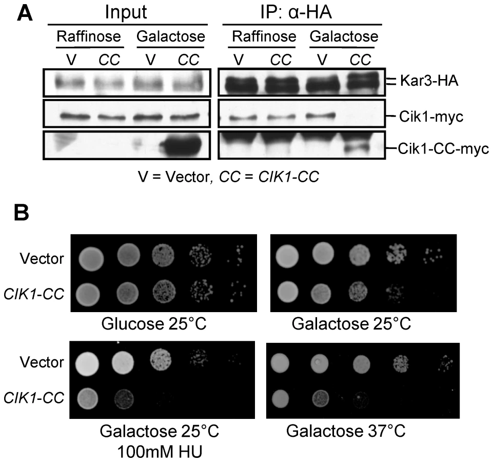
To further study the role of Cik1/Kar3 in chromosome bipolar attachment, we need to examine chromosome segregation in cik1Δ and kar3Δ mutants in the absence of the spindle checkpoint, which allows anaphase entry in spite of incorrect chromosome attachments. Because of the synthetic lethality, we have to develop a way to conditionally inactivate the Cik1/Kar3 complex. Kar3 and Cik1 associate with each other through their respective coiled-coil domains [32], thus overexpression of this domain may competitively disrupt the Cik1-Kar3 interaction. We constructed a plasmid PGALCIK1-CC that contains the coiled-coil domain of CIK1 under control of a galactose inducible promoter and the Cik1-Kar3 interaction in cells overexpressing CIK1-CC was examined.
Consistent with a previous report [28], we detected the interaction between Cik1 and Kar3 in control cells by co-immunoprecipitation. However, the Cik1-Kar3 interaction was completely abolished after CIK1-CC overexpression. Instead, the association of Kar3 with the coiled-coil domain of Cik1 (Cik1-CC) was detected, suggesting that the disruption of Cik1-Kar3 interaction by Cik1-CC is in a competitive manner (Figure 1A). Kar3 is able to form a heterodimer with either Cik1 or Vik1 [33], [34]. We also noticed that Vik1-Kar3 interaction was decreased in cells overexpressing CIK1-CC (Figure S1). Next we examined the phenotypes of cells overexpressing CIK1-CC and found that these cells grew slowly and were sensitive to HU. In addition, cells overexpressing CIK1-CC failed to grow at 37°C (Figure 1B), which is reminiscent of cik1Δ and kar3Δ mutants [29]. Therefore, we conclude that overexpression of CIK1-CC disrupts Cik1-Kar3 interaction and cells overexpressing CIK1-CC mimic the phenotypes of cik1Δ and kar3Δ mutants.
Tension checkpoint mutants abolish the anaphase entry delay induced by CIK1-CC overexpression
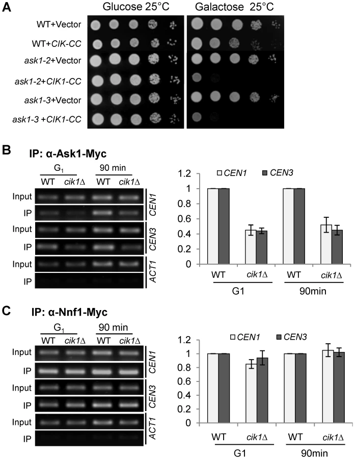
Next we examined the growth of the spindle checkpoint mutant mad1Δ and tension checkpoint mutants ipl1-321 and sgo1Δ after CIK1-CC overexpression. These mutant cells harboring a vector grew well on galactose plates, but the mutant cells with PGALCIK1-CC plasmids failed to grow on galactose plates (Figure 2A). Moreover, the checkpoint mutant cells lost viability shortly after the induction of CIK1-CC (Figure 2B). Surprisingly, ipl1-321 mutants with PGALCIK1-CC plasmids did not grow on galactose plates and lost viability after incubation in galactose medium at the permissive temperature 25°C (Figure 2A and 2B), which may be due to the fact that mutated Ipl1-321 protein shows significantly impaired kinase activity even at 25°C [35], [36].

We speculate that the slow growth phenotype in cells overexpressing CIK1-CC is attributed to the incorrect chromosome attachments that delay anaphase entry by stabilizing Pds1. Moreover, this delay likely depends on the spindle checkpoint. To test this idea, we compared the cell cycle progression in wild-type (WT), mad1Δ, sgo1Δ, and ipl1-321 cells after CIK1-CC overexpression. After G1 release into 25°C galactose medium for 200 min, 39% of WT cells with a control vector were large budded, while the percentage of large budded cells increased to 74% in those expressing CIK1-CC (Figure 2C). Consistent with the cell cycle delay, CIK1-CC overexpression also caused Pds1 protein stabilization. Strikingly, the cell cycle delay and Pds1 stabilization were eliminated not only in the spindle checkpoint mutant mad1Δ, but also in tension checkpoint mutants sgo1Δ and ipl1-321, which only detect lack of tension (Figure 2D). This result suggests that tension defects but not detached chromosomes activate the checkpoint in cells lacking Cik1/Kar3.
Cells overexpressing CIK1-CC exhibit a chromosome bipolar attachment defect
After the establishment of bipolar attachment, chromosomes congress to the spindle equator [37]. In budding yeast, the subsequent tension generation on chromosomes results in a transient separation of sister centromeres [38], [39]. cdc13-1 mutant cells arrest at preanaphase at high temperatures because of the activation of the DNA damage checkpoint and bipolar attachment is believed to be established in these arrested cells [40], [41], [42]. To assay the bipolar establishment in cells lacking Cik1/Kar3, we introduced the PGALCIK1-CC plasmid into cdc13-1 cells with GFP-marked centromeres of chromosome IV (CEN4-GFP) and mCherry-labeled microtubules (TUB1-mCherry). The relative localization of CEN4-GFP to the spindle was analyzed after G1 release into galactose medium at 32°C, the restrictive temperature for cdc13-1. Similar to cik1Δ mutant cells, overexpression of CIK1-CC also causes the formation of a dot-like spindle in some cells. Here, we only counted the cells with a metaphase spindle structure (>1.5 µM). After the establishment of bipolar attachment, we speculate that CEN4-GFP will either separate as two dots along the spindle or localize in the middle part of the spindle as a single dot. Nevertheless, the localization of a CEN4-GFP dot at the end of a spindle will suggest defective bipolar attachment. After G1 release for 150 min, only 12% of control cells showed a CEN4-GFP dot at one end of the spindle, but the percentage of cells with the GFP dot at the end of the spindle increased to 51% in those overexpressing CIK1-CC (Figure 3A), indicating that cells lacking Cik1/Kar3 function exhibit impaired chromosome bipolar attachment even when the spindle appears normal.

After tension establishment, kinetochores are resolved into two distinct clusters lying between the spindle poles before anaphase entry [43]. To test whether overexpression of CIK1-CC causes the failure of the formation of two kinetochore clusters, we introduced a PGALCIK1-CC plasmid into cdc13-1 strains with TUB1-mCherry and GFP-tagged MTW1, which encodes a kinetochore protein. After release from G1 into 32°C galactose medium for 150 min, 82% of cdc13-1 cells with a metaphase spindle showed two clearly resolved Mtw1-GFP foci in the absence of Cik1-CC induction. When CIK1-CC was overproduced, however, only 25% of cells exhibited two clear GFP foci among the cells with a normal metaphase spindle and many cells showed scattered GFP signals along the entire spindle (Figure 3B). Together, these results strongly suggest that dysfunctional Cik1/Kar3 leads to chromosome bipolar attachment defects. Although we cannot exclude the possibility that the abnormal spindle in cells lacking Cik1/Kar3 contributes to bipolar attachment defects, our data suggest a spindle-independent role of Cik1/Kar3 in bipolar attachment.
Cells with dysfunctional Cik1/Kar3 show syntelic attachment that allows chromosome mis-segregation in the absence of the spindle or the tension checkpoint
To further study the role of Cik1/Kar3 in chromosome bipolar attachment, we examined the chromosome segregation process in synchronized mad1Δ checkpoint mutant cells with CEN4-GFP TUB1-mCherry in the absence of Cik1/Kar3 function. After G1 release, CIK1-CC overexpression caused an obvious cell cycle delay in WT cells as indicated by the higher proportion of large budded cells, but mad1Δ suppressed this delay completely. Among the cells with an elongated spindle after G1 release for 150 min, more than 40% of mad1Δ cells showed CEN4-GFP co-segregation when Cik1-CC was overproduced, where one or two GFP dots were close to only one of the spindle poles. However, in mad1Δ cells with a vector control, no co-segregation of sister chromatids was observed (Figure 4A), indicating that Cik1-CC overproduction causes a kinetochore attachment defects.

The co-segregation of chromosome IV in mad1Δ mutant cells overexpressing CIK1-CC could be a consequence of syntelic attachment, monotelic attachment, or chromosome detachment. For a detached chromosome, both sister chromatids will stay in the mother cell after spindle elongation. For a chromosome with monotelic attachment, after anaphase onset the detached chromatid will stay in the mother cell but the attached one will move along with the connected spindle poles to either the mother or the daughter cell. Hence, it is impossible for both sister chromatids to move to the daughter cell together when a chromosome is either detached or with monotelic attachment. For a chromosome with syntelic attachment, however, the sister chromatids will co-segregate to either the mother or the daughter cell. Therefore, co-segregation of sister chromatids to the daughter cell will be an indication of syntelic attachment. We examined the frequency of sister-CEN4-GFP co-segregation into daughter cells in mad1Δ mutants overexpressing CIK1-CC. Mother cells are usually bigger in size and show a shmoo-like morphology because α-factor was used for G1 synchronization. Among the mad1Δ cells that show CEN4-GFP co-segregation after Cik1-CC induction, 45% of them have the GFP signal in the daughter cell (Figure 4A), indicating the presence of syntelic attachment. As this number is close to 50%, the chance of syntelic attachment to either spindle pole is similar.
If syntelic attachment in cells lacking Cik1/Kar3 function leads to a cell cycle delay, we expect that this delay depends on the tension checkpoint, because chromosomes with syntelic attachment are not under tension. Thus, we examined the chromosome segregation in ipl1-321 and sgo1Δ cells at 25°C after CIK1-CC overexpression. Strikingly, more than 40% ipl1-321 and sgo1Δ cells with an elongated spindle exhibited co-segregation of sister CEN4-GFPs after G1 release for 150 min, which is similar to mad1Δ checkpoint mutants. Among them, 54% of ipl1-321 and 48% of sgo1Δ cells showed exclusive daughter cell localization of CEN4-GFP signal. In contrast, no co-segregation was observed in the mutant cells with a vector control (Figure 4A). Given the fact that the loss of function of Ipl1 or Sgo1 fails to abolish the cell cycle arrest in response to detached chromosomes [19], [20], this result further indicates that dysfunctional Cik1/Kar3 induces syntelic attachment. Since we performed the experiments at 25°C, the data demonstrate that ipl1-321 mutant cells lose tension checkpoint function at the permissive temperature, which is in agreement with the observation that ipl1-321 mutants exhibit reduced kinase activity at the permissive temperature [36].
We also used a live-cell imaging system to follow chromosome segregation in sgo1Δ mutants overexpressing CIK1-CC in order to clarify if some sister chromatids are connected to a single spindle pole. When incubated in galactose medium, the sister CEN4-GFPs migrated along with one single spindle pole and entered the daughter cell as the spindle elongated in the cell shown in Figure 4B and Video S1, indicating that both sister chromatids of chromosome IV are connected to one spindle pole. During spindle elongation, we observed separation of the two GFP dots at some time points, suggesting the absence of sister chromatid cohesion. Therefore, the sister chromatid co-segregation observed in this representative cell is likely a consequence of syntelic attachment, but not monotelic attachment. Chromosome mis-segregation was not observed in WT cells overexpressing CIK1-CC in this live-cell assay.
Residual cohesion may contribute to sister-chromatid co-segregation of a chromosome with monotelic attachment. To further distinguish syntelic from monotelic attachment, we examined sister-chromatid segregation in mcd1-1 cohesin mutants while overexpressing CIK1-CC. When incubated at 37°C, the absence of cohesion in mcd1-1 mutants not only abolishes the connection between sister chromatids, but also allows the spindle to elongate regardless of checkpoint activation [44]. We introduced a vector and a PGALCIK1-CC plasmid into a mcd1-1 CEN5-GFP TUB1-mCherry strain. After G1 release into galactose medium at 37°C for 200 min, 31% of mcd1-1 mutant cells with an elongated spindle exhibited co-segregation of sister CEN5-GFPs when Cik1-CC was induced. In the absence of Cik1-CC expression, however, only 4% mcd1-1 cells with an elongated spindle showed CEN5-GFP co-segregation, presumably because cohesion also contributes to bipolar attachment [45] (Figure 4C). Moreover, the chance of sister-chromatid co-segregation into the mother or the daughter cell is similar. The dramatically increased frequency of sister-chromatid co-segregation in cohesin mutants after Cik1-CC overexpression further demonstrates the presence of syntelic attachment. Together, these data support the conclusion that loss of function of Cik1/Kar3 by overexpressing CIK1-CC causes syntelic attachment, where two sister kinetochores attach to the same spindle pole.
A temperature sensitive mutant kar3-64 shows chromosomes with syntelic attachment
Overexpression of the coiled-coil domain of Cik1 may disrupt the function of other proteins with a coiled-coil domain, which could also contribute to syntelic attachments. To exclude this possibility, we examined chromosome segregation in a temperature sensitive kar3-64 mutant in the absence of Sgo1. The mutated Kar3-64 protein loses its function at high temperature because kar3-64 is synthetically lethal with kip3Δ at 35°C but not at room temperature [46]. WT, sgo1Δ, kar3-64, and kar3-64 sgo1Δ strains with CEN4-GFP TUB1-mCherry were first arrested in G1 phase and then released into 35°C medium to inactivate Kar3. The accumulation of large budded cells in kar3-64 mutants indicates the loss of Kar3 function (Figure 5A). Similar to the cells overexpressing CIK1-CC, the cell cycle delay in kar3-64 mutant was abolished by sgo1Δ. We also found that about 40% of kar3-64 sgo1Δ double mutant cells with an elongated spindle showed CEN4-GFP co-segregation after release for 120 and 150 min at 35°C (Figure 5B), which is comparable to sgo1Δ cells overexpressing CIK1-CC. The majority of kar3-64 sgo1Δ mutant cells exited mitosis after release for 180 min. At this time point, 38% of the G1 cells were either absent for CEN4-GFP signal or showed two CEN4-GFP dots, suggesting the gain or loss of chromosome IV after mitosis (Figure 5C). It is also possible that some G1 cells have two CEN4-GFP dots but they are too close to be distinguished by microscopy. Consistently, only 18% of kar3-64 sgo1Δ mutants were viable after G1 release for 180 min, but 95% of WT and sgo1Δ cells as well as 61% of kar3-64 cells were viable (Figure 5D). kar3-64 cells exhibited partial viability loss presumably due to the inability to recover from mitotic arrest. These results validate the conclusion that the loss of function of Kar3 causes syntelic attachment.

The bipolar attachment defects in kar3 mutants or in cells overexpressing CIK1-CC could be a result of dysfunctional Cik1/Kar3 or Vik1/Kar3, because overexpression of CIK1-CC also partially disrupts Vik1-Kar3 interactions (Figure S1). Moreover, previous observation that vik1Δ mutant is synthetically lethal with ipl1-321 indicates a possible role of Vik1 in chromosome segregation [45]. To test whether dysfunctional Vik1 also contributes to syntelic attachment, we examined the establishment of bipolar attachment in cdc13-1 vik1Δ cells. Like cdc13-1 single mutant, more than 80% of cdc13-1 vik1Δ cells showed either separated CEN4-GFP dots or one GFP dot at the center region of the spindle after G1 release for 90 min (Figure S2). We further examined the segregation of sister chromatids in vik1Δ mad1Δ double mutant cells and no mis-segregation was observed. We also crossed vik1Δ with ipl1-321 and sgo1Δ. Surprisingly, we obtained vik1Δ ipl1-321 and vik1Δ sgo1Δ double mutants and these mutants did not show co-segregation of sister chromatids (Figure S3). Therefore, vik1Δ mutants exhibit distinct phenotypes from cik1Δ. It is likely that only the Cik1/Kar3 complex is required for the establishment of chromosome bipolar attachment.
Cik1 mediates Kar3–kinetochore interaction
Cik1 and Vik1 are the two Kar3 partners in budding yeast and Cik1/Kar3 localizes along the length of the spindle, and probably at interpolar microtubule plus ends [25], [33]. Kar3 was also found to bind to the kinetochore to promote its transport along astral microtubules towards spindle poles [26]. The similar mitotic defects in cik1 and kar3 mutants suggest that Cik1 likely mediates the association of Kar3 with kinetochores. Based on the genome-wide yeast two-hybrid assay, Kar3 was shown to interact with Nnf1, a kinetochore protein in the MIND complex, [7], [47], [48]. Using a co-immunoprecipitation (co-IP) approach we found that Nnf1-myc was able to pull down Kar3-HA, and vice versa, confirming the Kar3-kinetochore interaction in vivo, although it remains inconclusive whether this Kar3-Nnf1 interaction is direct (Figure 6A). Then, we examined Kar3-Nnf1 interaction in the absence of either Cik1 or Vik1. As shown in Figure 6B, deletion of CIK1 but not VIK1 abolished this interaction completely, suggesting that the Kar3-kinetochore interaction is dependent on Cik1. Interestingly, this interaction was obviously increased in vik1Δ mutant cells, presumably because more Kar3 protein is available for the binding to kinetochores. Consistently, chromatin immunoprecipitation (ChIP) data shows diminished Kar3-centromere association in cik1Δ cells (Figure 6C). If Cik1 mediates Kar3-Nnf1 interaction, the overexpression of the coiled-coil domain of Cik1 should disrupt this interaction, because CIK1-CC overexpression disrupts Cik1-Kar3 interaction (Figure 1A). Indeed, Kar3-HA failed to pull down Nnf1-myc in cells overexpressing CIK1-CC (Figure 6D).

Data from the Sorger lab suggest that Kar3 may associate with detached kinetochores [43]. Moreover, Kar3 is essential for the lateral sliding of chromosomes towards spindle poles during S-phase [27]. Therefore, the association of Kar3 with kinetochores might be cell cycle regulated. To test this possibility, we compared Kar3-Nnf1 interaction in different cell cycle stages. Kar3 interacted with Nnf1 in both HU - and nocodazole-arrested cells (Figure 6E), when bipolar attachment has not established yet. cdc13-1 mutant cells arrest at preanaphase with established bipolar attachment [40]. Interestingly, the Kar3-Nnf1 interaction was not detected in cdc13-1 mutant cells after 2 hr incubation at 32°C (Figure 6F). Similarly, Kar3 did not associate with Nnf1 in cdc15-2-arrested telophase cells. The decreased Kar3-Nnf1 interaction in cdc13-1 or cdc15-2 mutant cells could be due to the degradation of Cik1 [49], thus the Cik1 protein levels were examined in WT, cdc13-1, and cdc15-2 mutant cells incubated at permissive or no-permissive temperatures. It is clear that the mutant cells exhibit Cik1 protein levels comparable to WT cells when incubated at high temperatures (Figure 6G). The results suggest that the Cik1/Kar3 complex associates with kinetochores before the establishment of bipolar attachment. This association might be essential for chromosome transport as well as the achievement of bipolar attachment, but lack of this mechanism will contribute to syntelic attachment.
The Cik1/Kar3 complex is required for efficient DASH–centromere binding
The DASH kinetochore complex contains 10 protein subunits including Dam1 and Ask1. Unlike other kinetochore proteins, the association of the DASH complex with kinetochores occurs only after kinetochore-microtubule interaction [2]. Interestingly, kar3Δ has been shown to be synthetically lethal with dam1-1 mutant [50]. We found that overexpression of CIK1-CC caused lethality to ask1-2 and ask1-3 mutants (Figure 7A). One possibility is that the Cik1/Kar3 complex promotes bipolar attachment by inducing the DASH-kinetochore interaction. Therefore, we performed ChIP assays to examine DASH-centromere interaction in synchronous cell cultures. cdc13-1 mutants were used to arrest cells at preanaphase, when DASH complexes have already been loaded onto centromeres [2]. Interestingly, cdc13-1 cik1Δ cells exhibited reduced Ask1-centromere interaction in G1 phase as well as after G1 release for 90 min (Figure 7B). In order to determine whether the Ask1 binding defect is a result of impaired kinetochore integrity, we also compared the centromere binding of another kinetochore protein Nnf1. In contrast to Ask1, the association of Nnf1 with centromeric DNA was similar in synchronous cdc13-1 and cdc13-1 cik1Δ cells either before or after G1 release (Figure 7C), indicating that the core kinetochore structure is intact. These results suggest that the Cik1/Kar3 complex facilitates the association of the DASH complex with the core kinetochore proteins. It is our future interest to determine if decreased DASH-kinetochore interaction is the cause or a consequence of syntelic attachments.
Discussion
In budding yeast, Kar3 is the only kinesin with minus-end-directed motor activity and the interaction with its partners, Cik1 and Vik1, is essential for its motor activity. Our data clearly demonstrate the presence of syntelic attachments in cells lacking Cik1/Kar3 function based on the following observations: first, in the absence of the spindle checkpoint, the loss of function of Cik1/Kar3 induced significant sister-chromatid co-segregation into the daughter cell. Moreover, a dysfunctional tension checkpoint is sufficient to enable anaphase entry as well as co-segregation of sister chromatids in cells lacking Cik1/Kar3 function. Because it is well established that cells with tensionless chromosomes require the tension checkpoint for cell cycle arrest, this result indicates the presence of chromosomes with syntelic attachment in cells lacking functional Cik1/Kar3. Furthermore, our live-cell imaging data show the migration of both sister chromatids along with one spindle pole during anaphase in sgo1Δ cells overexpressing CIK1-CC. As recent evidence indicates that residual sister chromatid cohesion remains during early anaphase [51], one can argue that the sister chromatid co-segregation is a consequence of monotelic attachment, where only one of the sister kinetochores connects to the spindle pole but the other co-migrates with the sister because of the residual cohesion. Our data suggest that this scenario is unlikely, because we detected separated CEN4-GFP dots during anaphase in a sgo1Δ cell overexpressing CIK1-CC, indicating the absence of cohesion. In addition, we found that dysfunctional Cik1/Kar3 also induces sister chromatid co-segregation in cohesin mutant cells. Therefore, we conclude that inactivation of the Cik1/Kar3 complex induces syntelic attachment.
In cells overexpressing the coiled-coil domain of CIK1, we observed delayed Pds1 degradation, indicating the activation of the spindle checkpoint. However, either ipl1-321 or sgo1Δ abolished this delay completely. In addition, both ipl1-321 and sgo1Δ mutants show normal timing of spindle elongation when CIK1-CC is overexpressed, resulting in a high frequency of sister-chromatid co-segregation that is comparable to mad1Δ mutants. Previous data indicate that ipl1 mutants, but not sgo1, generate detached chromosomes when these chromosomes are not under tension [22], thus it is speculated that Ipl1 activates the spindle checkpoint by generating detached chromosomes when tension is absent. However, the complete loss of checkpoint function in sgo1 mutant cells overexpressing CIK1-CC suggests that the generation of detached chromosomes is not necessary to activate the spindle checkpoint in the presence of syntelic attachments. Interestingly, we found that ipl1-321 mutants show checkpoint defect even when grown at 25°C. Therefore, the decreased kinase activity in ipl1-321 mutants at the permissive temperature may be unable to execute the checkpoint function, but is sufficient to support normal chromosome segregation.
Although our data indicate that cells lacking functional Cik1/Kar3 show syntelic attachments, the molecular function of this motor complex in chromosome bipolar attachment remains elusive. One possible explanation is that the Kar3-dependent poleward chromosome movement facilitates chromosome bipolar attachment. After the initial chromosome capture, Cik1/Kar3-mediated sliding of this chromosome along spindle microtubules will orient sister kinetochores so that the detached kinetochore will face the opposite spindle pole, thereby facilitating bipolar attachment. In agreement with this possibility, we found that Cik1 mediates the association of Kar3 with kinetochores and this association only occurs before chromosome bipolar attachment. In mammals and flies, kinetochore dynein mediates the poleward chromosome movement, which may also facilitate the correct orientation of sister kinetochores through a similar mechanism [52], [53]. Another possibility is that the abnormal spindle structure in cik1 and kar3 mutant cells may contribute to the high frequency of syntelic attachments. At 37°C, cik1Δ and kar3Δ mutants arrest with a dot-like spindle structure and many mutant cells show spindle defects even when incubated at room temperature [23], [29], [33]. We examined chromosome bipolar attachment in cells arrested at preanaphase and found that the disruption of Cik1/Kar3 function by overexpressing CIK1-CC increases the chance of co-localization of CEN4-GFP with one spindle pole in cells with a metaphase spindle that appears normal. This result suggests that the bipolar attachment defect in cells lacking Cik1/Kar3 function could be independent of the spindle defect. To separate the spindle and kinetochore functions of Cik1/Kar3, we need to identify the kinetochore protein that directly binds to the Cik1/Kar3 complex and define the domain responsible for this interaction. Mutation of this domain may selectively disrupt the kinetochore function of Cik1/Kar3 but maintain its spindle function.
In mammalian cells, syntelic attachment can be induced by treatment with a small molecule monastrol, which inhibits the activity of a kinesin Eg5 [54], [55]. Moreover, the treatment of mammalian cells with a low-dose of taxol will prevent tension generation on chromosomes [56]. In budding yeast, two methods have been widely used to introduce a tension defects for the study of the tension sensing mechanism. One is to completely block DNA replication where the absence of sister chromatids prevents tension generation, while another method is to abolish sister chromatid cohesion by using temperature sensitive mcd1/scc1 mutants or by expressing MCD1 from a galactose inducible promoter [19], [20]. The disadvantage of these methods is that Pds1 protein level is the only marker to monitor the checkpoint activity. Because the lack of sister-chromatid cohesion allows the spindle to elongate regardless of checkpoint activation, we cannot use spindle elongation to monitor anaphase entry. Moreover it is difficult to analyze the role of the tension checkpoint in faithful chromosome segregation. Another disadvantage is that these methods will kill the cells after the induction of tension defects, thus it is impossible to perform a genetic screen for additional genes that are required for survival in the presence of tension defects. Here we report a new approach to induce syntelic attachments by inactivating the Cik1/Kar3 motor complex, which prevents tension generation on chromosomes but maintains intact kinetochore attachment. We have demonstrated that overexpression of the coiled-coil domain of Cik1 from a GAL promoter disrupts Cik1-Kar3 interaction, which allows us to conditionally induce syntelic attachment by growing cells with PGALCIK1-CC plasmid in galactose medium. This approach will be a critical tool to study the response to tension defects in budding yeast.
Materials and Methods
Strains, plasmids, and growth conditions
The strains used in this study are derivatives of W303 and listed in Table S1. Gene deletions and epitope tagging were performed by using a PCR-based protocol [57]. The PGALCIK1-CC plasmid was constructed by inserting the CIK1 coiled-coil fragment into a CEN-TRP-GAL-myc vector.
To arrest yeast cells in G1 phase, 5 µg/ml α-factor was added into mid-log phase cells grown in YPD or in TRP dropout medium containing 2% raffinose at 25°C for 2.5 hr. G1-arrested cells were centrifuged and washed twice with water to release into YPD at 32°C for cdc13-1 arrest or TRP dropout medium containing 2% galactose at 25°C for CIK1-CC overexpression. To block the next cell cycle, 15 µg/ml α-factor was added when majority of the cells were budded. Hydroxyurea was purchased from ACROS Organics and the final concentration was 100 mM for HU plates.
Protein techniques
The yeast protein samples were separated and detected as described previously [58]. Protein samples were prepared using an alkaline method and were resolved by 10% SDS - PAGE. Primary antibodies (anti-myc and anti-HA) were purchased from Covance (Madison, WI), and anti-Pgk1 antibody was from Molecular Probes (Eugene, OR). The HRP-conjugated secondary antibody was purchased from Jackson ImmunoResearch (West Grove, PA).
Fluorescence microcopy
Cells were collected and fixed with 3.7% formaldehyde for 15 min at room temperature. The cells were washed once with 1×PBS (pH7.2) and then resuspended in 1×PBS buffer to examine fluorescence signals with a microscope (Zeiss Axioplan 2).
Co-immunoprecipitation (co-IP) and chromatin immunoprecipitation (ChIP) assay
Cell cultures were collected and washed once with water. After being resuspended in RIPA buffer (25 mM Tris PH7.5, 10 mM EDTA, 150 mM NaCl and 0.05% Tween-20) supplied with protease inhibitors, cells were homogenized with a bead-beater. The resulting cell extracts were incubated with primary antibody overnight at 4°C. The cell extracts were then incubated with protein-A conjugated agarose beads (Santa Cruz Biotechnology), which was pre-incubated with BSA at 4°C. After incubation for 1 hr, the beads were collected by centrifugation and washed with RIPA buffer for three times. Equal volume RIPA and protein loading buffer were added and the protein samples were boiled for 5 min for Western blot analysis. The ChIP assay was performed as described previously [59].
Live-cell fluorescence microscopy
For live-cell microscopy, we used a concave glass slide as a culture chamber, which was filled with 2% agarose dissolved in galactose medium. The agarose pad was solidified for 5 min at room temperature before use. Cells were first arrested in G1 phase in raffinose medium. After release into galactose medium for 2 hr, 1.5 µl concentrated cells were laid on the top of the agarose pad, which was then sealed with a piece of cover glass. Live-cell microscopy was carried out on a DeltaVision imaging system equipped with an environmental chamber (Applied Precision, Inc.). All live-cell images were acquired at 25°C with a 100× (NA = 1.41) objective lens on an Olympus ix71 microscope. A total of 8 z-stacks were collected at each time point and each optical section was 0.5 µm thick. Exposure time for each optical section was set between 60 and 100 ms and the time-lapse interval was set at 2 min. Projected images were used for display.
Supporting Information
Zdroje
1. PinskyBABigginsS 2005 The spindle checkpoint: tension versus attachment. Trends Cell Biol 15 486 493
2. LiYBachantJAlcasabasAAWangYQinJ 2002 The mitotic spindle is required for loading of the DASH complex onto the kinetochore. Genes Dev 16 183 197
3. WestermannSAvila-SakarAWangHWNiederstrasserHWongJ 2005 Formation of a dynamic kinetochore - microtubule interface through assembly of the Dam1 ring complex. Mol Cell 17 277 290
4. WestermannSWangHWAvila-SakarADrubinDGNogalesE 2006 The Dam1 kinetochore ring complex moves processively on depolymerizing microtubule ends. Nature 440 565 569
5. JankeCOrtizJTanakaTULechnerJSchiebelE 2002 Four new subunits of the Dam1-Duo1 complex reveal novel functions in sister kinetochore biorientation. EMBO J 21 181 193
6. WestermannSDrubinDGBarnesG 2007 Structures and Functions of Yeast Kinetochore Complexes. Annu Rev Biochem
7. De WulfPMcAinshADSorgerPK 2003 Hierarchical assembly of the budding yeast kinetochore from multiple subcomplexes. Genes Dev 17 2902 2921
8. HoytMATotisLRobertsBT 1991 S. cerevisiae genes required for cell cycle arrest in response to loss of microtubule function. Cell 66 507 517
9. LiRMurrayAW 1991 Feedback control of mitosis in budding yeast. Cell 66 519 531
10. HardwickKGWeissELucaFCWineyMMurrayAW 1996 Activation of the budding yeast spindle assembly checkpoint without mitotic spindle disruption. Science 273 953 956
11. WangYBurkeDJ 1995 Checkpoint genes required to delay cell division in response to nocodazole respond to impaired kinetochore function in the yeast Saccharomyces cerevisiae. Mol Cell Biol 15 6838 6844
12. HardwickKGJohnstonRCSmithDLMurrayAW 2000 MAD3 encodes a novel component of the spindle checkpoint which interacts with Bub3p, Cdc20p, and Mad2p. J Cell Biol 148 871 882
13. ChenRH 2002 BubR1 is essential for kinetochore localization of other spindle checkpoint proteins and its phosphorylation requires Mad1. J Cell Biol 158 487 496
14. Cohen-FixOPetersJMKirschnerMWKoshlandD 1996 Anaphase initiation in Saccharomyces cerevisiae is controlled by the APC-dependent degradation of the anaphase inhibitor Pds1p. Genes Dev 10 3081 3093
15. CioskRZachariaeWMichaelisCShevchenkoAMannM 1998 An ESP1/PDS1 complex regulates loss of sister chromatid cohesion at the metaphase to anaphase transition in yeast. Cell 93 1067 1076
16. UhlmannFLottspeichFNasmythK 1999 Sister-chromatid separation at anaphase onset is promoted by cleavage of the cohesin subunit Scc1. Nature 400 37 42
17. LiXNicklasRB 1995 Mitotic forces control a cell-cycle checkpoint. Nature 373 630 632
18. KeatingPRachidiNTanakaTUStarkMJ 2009 Ipl1-dependent phosphorylation of Dam1 is reduced by tension applied on kinetochores. J Cell Sci 122 4375 4382
19. BigginsSMurrayAW 2001 The budding yeast protein kinase Ipl1/Aurora allows the absence of tension to activate the spindle checkpoint. Genes Dev 15 3118 3129
20. IndjeianVBSternBMMurrayAW 2005 The centromeric protein Sgo1 is required to sense lack of tension on mitotic chromosomes. Science 307 130 133
21. TanakaTURachidiNJankeCPereiraGGalovaM 2002 Evidence that the Ipl1-Sli15 (Aurora kinase-INCENP) complex promotes chromosome bi-orientation by altering kinetochore-spindle pole connections. Cell 108 317 329
22. PinskyBAKungCShokatKMBigginsS 2006 The Ipl1-Aurora protein kinase activates the spindle checkpoint by creating unattached kinetochores. Nat Cell Biol 8 78 83
23. MeluhPBRoseMD 1990 KAR3, a kinesin-related gene required for yeast nuclear fusion. Cell 60 1029 1041
24. EndowSAKangSJSatterwhiteLLRoseMDSkeenVP 1994 Yeast Kar3 is a minus-end microtubule motor protein that destabilizes microtubules preferentially at the minus ends. EMBO J 13 2708 2713
25. GardnerMKHaaseJMythreyeKMolkJNAndersonM 2008 The microtubule-based motor Kar3 and plus end-binding protein Bim1 provide structural support for the anaphase spindle. J Cell Biol 180 91 100
26. TanakaKMukaeNDewarHvan BreugelMJamesEK 2005 Molecular mechanisms of kinetochore capture by spindle microtubules. Nature 434 987 994
27. TanakaKKitamuraEKitamuraYTanakaTU 2007 Molecular mechanisms of microtubule-dependent kinetochore transport toward spindle poles. J Cell Biol 178 269 281
28. PageBDSnyderM 1992 CIK1: a developmentally regulated spindle pole body-associated protein important for microtubule functions in Saccharomyces cerevisiae. Genes Dev 6 1414 1429
29. LiuHJinFLiangFTianXWangY 2011 The Cik1/Kar3 motor complex is required for the proper kinetochore-microtubule interaction after stressful DNA replication. Genetics 187 397 407
30. DanielJAKeyesBENgYPFreemanCOBurkeDJ 2006 Diverse functions of spindle assembly checkpoint genes in Saccharomyces cerevisiae. Genetics 172 53 65
31. TongAHLesageGBaderGDDingHXuH 2004 Global mapping of the yeast genetic interaction network. Science 303 808 813
32. BarrettJGManningBDSnyderM 2000 The Kar3p kinesin-related protein forms a novel heterodimeric structure with its associated protein Cik1p. Mol Biol Cell 11 2373 2385
33. ManningBDBarrettJGWallaceJAGranokHSnyderM 1999 Differential regulation of the Kar3p kinesin-related protein by two associated proteins, Cik1p and Vik1p. J Cell Biol 144 1219 1233
34. SproulLRAndersonDJMackeyATSaundersWSGilbertSP 2005 Cik1 targets the minus-end kinesin depolymerase kar3 to microtubule plus ends. Curr Biol 15 1420 1427
35. KotwaliwaleCVFreiSBSternBMBigginsS 2007 A pathway containing the Ipl1/aurora protein kinase and the spindle midzone protein Ase1 regulates yeast spindle assembly. Dev Cell 13 433 445
36. MakrantoniVStarkMJ 2009 Efficient chromosome biorientation and the tension checkpoint in Saccharomyces cerevisiae both require Bir1. Mol Cell Biol 29 4552 4562
37. GardnerMKBouckDCPaliulisLVMeehlJBO'TooleET 2008 Chromosome congression by Kinesin-5 motor-mediated disassembly of longer kinetochore microtubules. Cell 135 894 906
38. HeXAsthanaSSorgerPK 2000 Transient sister chromatid separation and elastic deformation of chromosomes during mitosis in budding yeast. Cell 101 763 775
39. GoshimaGYanagidaM 2000 Establishing biorientation occurs with precocious separation of the sister kinetochores, but not the arms, in the early spindle of budding yeast. Cell 100 619 633
40. BachantJAlcasabasABlatYKlecknerNElledgeSJ 2002 The SUMO-1 isopeptidase Smt4 is linked to centromeric cohesion through SUMO-1 modification of DNA topoisomerase II. Mol Cell 9 1169 1182
41. TangXWangY 2006 Pds1/Esp1-dependent and -independent sister chromatid separation in mutants defective for protein phosphatase 2A. Proc Natl Acad Sci U S A 103 16290 16295
42. LiangFWangY 2007 DNA damage checkpoints inhibit mitotic exit by two different mechanisms. Mol Cell Biol 27 5067 5078
43. TytellJDSorgerPK 2006 Analysis of kinesin motor function at budding yeast kinetochores. J Cell Biol 172 861 874
44. GuacciVKoshlandDStrunnikovA 1997 A direct link between sister chromatid cohesion and chromosome condensation revealed through the analysis of MCD1 in S. cerevisiae. Cell 91 47 57
45. NgTMWaplesWGLavoieBDBigginsS 2009 Pericentromeric sister chromatid cohesion promotes kinetochore biorientation. Mol Biol Cell 20 3818 3827
46. CottinghamFRGheberLMillerDLHoytMA 1999 Novel roles for Saccharomyces cerevisiae mitotic spindle motors. J Cell Biol 147 335 350
47. NewmanJRWolfEKimPS 2000 A computationally directed screen identifying interacting coiled coils from Saccharomyces cerevisiae. Proc Natl Acad Sci U S A 97 13203 13208
48. WongJNakajimaYWestermannSShangCKangJS 2007 A protein interaction map of the mitotic spindle. Mol Biol Cell 18 3800 3809
49. BenantiJAMatyskielaMEMorganDOToczyskiDP 2009 Functionally distinct isoforms of Cik1 are differentially regulated by APC/C-mediated proteolysis. Mol Cell 33 581 590
50. JonesMHBachantJBCastilloARGiddingsTHJrWineyM 1999 Yeast Dam1p is required to maintain spindle integrity during mitosis and interacts with the Mps1p kinase. Mol Biol Cell 10 2377 2391
51. RenshawMJWardJJKanemakiMNatsumeKNedelecFJ 2010 Condensins promote chromosome recoiling during early anaphase to complete sister chromatid separation. Dev Cell 19 232 244
52. SharpDJRogersGCScholeyJM 2000 Cytoplasmic dynein is required for poleward chromosome movement during mitosis in Drosophila embryos. Nat Cell Biol 2 922 930
53. BaderJRVaughanKT 2010 Dynein at the kinetochore: Timing, Interactions and Functions. Semin Cell Dev Biol 21 269 275
54. MayerTUKapoorTMHaggartySJKingRWSchreiberSL 1999 Small molecule inhibitor of mitotic spindle bipolarity identified in a phenotype-based screen. Science 286 971 974
55. KapoorTMMayerTUCoughlinMLMitchisonTJ 2000 Probing spindle assembly mechanisms with monastrol, a small molecule inhibitor of the mitotic kinesin, Eg5. J Cell Biol 150 975 988
56. FamulskiJKChanGK 2007 Aurora B kinase-dependent recruitment of hZW10 and hROD to tensionless kinetochores. Curr Biol 17 2143 2149
57. LongtineMSMcKenzieA3rdDemariniDJShahNGWachA 1998 Additional modules for versatile and economical PCR-based gene deletion and modification in Saccharomyces cerevisiae. Yeast 14 953 961
58. LiuHWangY 2006 The function and regulation of budding yeast Swe1 in response to interrupted DNA synthesis. Mol Biol Cell 17 2746 2756
59. LiuHLiangFJinFWangY 2008 The coordination of centromere replication, spindle formation, and kinetochore-microtubule interaction in budding yeast. PLoS Genet 4 e1000262 doi:10.1371/journal.pgen.1000262
Štítky
Genetika Reprodukční medicínaČlánek vyšel v časopise
PLOS Genetics
2012 Číslo 2
Nejčtenější v tomto čísle
- Gene Expression and Stress Response Mediated by the Epigenetic Regulation of a Transposable Element Small RNA
- Contrasting Properties of Gene-Specific Regulatory, Coding, and Copy Number Mutations in : Frequency, Effects, and Dominance
- Homeobox Genes Critically Regulate Embryo Implantation by Controlling Paracrine Signaling between Uterine Stroma and Epithelium
- Nondisjunction of a Single Chromosome Leads to Breakage and Activation of DNA Damage Checkpoint in G2