-
Články
Reklama
- Vzdělávání
- Časopisy
Top články
Nové číslo
- Témata
Reklama- Videa
- Podcasty
Nové podcasty
Reklama- Kariéra
Doporučené pozice
Reklama- Praxe
ReklamaA Novel Unstable Duplication Upstream of Predisposes to a Breed-Defining Skin Phenotype and a Periodic Fever Syndrome in Chinese Shar-Pei Dogs
Hereditary periodic fever syndromes are characterized by recurrent episodes of fever and inflammation with no known pathogenic or autoimmune cause. In humans, several genes have been implicated in this group of diseases, but the majority of cases remain unexplained. A similar periodic fever syndrome is relatively frequent in the Chinese Shar-Pei breed of dogs. In the western world, Shar-Pei have been strongly selected for a distinctive thick and heavily folded skin. In this study, a mutation affecting both these traits was identified. Using genome-wide SNP analysis of Shar-Pei and other breeds, the strongest signal of a breed-specific selective sweep was located on chromosome 13. The same region also harbored the strongest genome-wide association (GWA) signal for susceptibility to the periodic fever syndrome (praw = 2.3×10⁻⁶, pgenome = 0.01). Dense targeted resequencing revealed two partially overlapping duplications, 14.3 Kb and 16.1 Kb in size, unique to Shar-Pei and upstream of the Hyaluronic Acid Synthase 2 (HAS2) gene. HAS2 encodes the rate-limiting enzyme synthesizing hyaluronan (HA), a major component of the skin. HA is up-regulated and accumulates in the thickened skin of Shar-Pei. A high copy number of the 16.1 Kb duplication was associated with an increased expression of HAS2 as well as the periodic fever syndrome (p<0.0001). When fragmented, HA can act as a trigger of the innate immune system and stimulate sterile fever and inflammation. The strong selection for the skin phenotype therefore appears to enrich for a pleiotropic mutation predisposing these dogs to a periodic fever syndrome. The identification of HA as a major risk factor for this canine disease raises the potential of this glycosaminoglycan as a risk factor for human periodic fevers and as an important driver of chronic inflammation.
Published in the journal: . PLoS Genet 7(3): e32767. doi:10.1371/journal.pgen.1001332
Category: Research Article
doi: https://doi.org/10.1371/journal.pgen.1001332Summary
Hereditary periodic fever syndromes are characterized by recurrent episodes of fever and inflammation with no known pathogenic or autoimmune cause. In humans, several genes have been implicated in this group of diseases, but the majority of cases remain unexplained. A similar periodic fever syndrome is relatively frequent in the Chinese Shar-Pei breed of dogs. In the western world, Shar-Pei have been strongly selected for a distinctive thick and heavily folded skin. In this study, a mutation affecting both these traits was identified. Using genome-wide SNP analysis of Shar-Pei and other breeds, the strongest signal of a breed-specific selective sweep was located on chromosome 13. The same region also harbored the strongest genome-wide association (GWA) signal for susceptibility to the periodic fever syndrome (praw = 2.3×10⁻⁶, pgenome = 0.01). Dense targeted resequencing revealed two partially overlapping duplications, 14.3 Kb and 16.1 Kb in size, unique to Shar-Pei and upstream of the Hyaluronic Acid Synthase 2 (HAS2) gene. HAS2 encodes the rate-limiting enzyme synthesizing hyaluronan (HA), a major component of the skin. HA is up-regulated and accumulates in the thickened skin of Shar-Pei. A high copy number of the 16.1 Kb duplication was associated with an increased expression of HAS2 as well as the periodic fever syndrome (p<0.0001). When fragmented, HA can act as a trigger of the innate immune system and stimulate sterile fever and inflammation. The strong selection for the skin phenotype therefore appears to enrich for a pleiotropic mutation predisposing these dogs to a periodic fever syndrome. The identification of HA as a major risk factor for this canine disease raises the potential of this glycosaminoglycan as a risk factor for human periodic fevers and as an important driver of chronic inflammation.
Introduction
Shar-Pei dogs have been companion animals for centuries within China where they were commissioned to guard and hunt, and to sometimes serve as fighting animals. At the beginning of the communist era dog ownership was highly taxed and the breed was brought close to extinction. A few Chinese Shar-Pei dogs were exported to the United States in the early 1970's and Shar-Pei descending from this limited number of animals have undergone strong selection for a wrinkled skin phenotype and heavily padded muzzle and are called the “meatmouth” type (Figure 1A–1C) and have now found global popularity. The ancestral Shar-Pei, referred to as the “traditional” type Shar-Pei, still occurs and it presents with a less accentuated skin condition (Figure 1D). The major constituent of the deposit in the thickened skin is hyaluronan or hyaluronic acid (HA). HA is a large, multifunctional, linear, negatively charged, non-sulfated glycosaminoglycan of the extracellular and pericellular matrices. It is composed of repeating disaccharides and is widely spread throughout epithelial, connective and neural tissues [1], [2]. The biological role of HA depends on its size, location and equilibrium between synthesis and degradation [1]–[3]. Meatmouth Shar-Pei show two - to five-fold higher serum levels of HA compared to other breeds [4], allowing us to propose the term hyaluronanosis, a definition also used for a comparable human condition [5]. HA is synthesized at the plasma membrane by three HA synthases, HAS1, HAS2 and HAS3, with HAS2 being the rate limiting-enzyme [6]. HAS2 is overexpressed in dermal fibroblasts of Shar-Pei compared with other canine breeds [7] suggesting a regulatory mutation as causative for hyaluronanosis. HA is deposited throughout the skin of Shar-Pei, often in microscopic lakes and grossly evident vesicles, leading to the formation of thickened skin folds around the head and tibiotarsal (hock) joints (Figure 1E). Almost all Shar-Pei seem to be affected by hyaluronanosis, however the extent varies among individuals and adults exhibit less skin folds and hyaluronanosis than puppies. Strong selection by breeders for dogs who retained their skin folds into adulthood has altered the phenotype of the breed to the more commonly heavily wrinkled meatmouth type.
Fig. 1. The phenotypic spectrum of the Chinese Shar-Pei. 
Following strong selection for the “wrinkled” skin phenotype, Shar-Pei dogs in the western world most commonly present as the meatmouth type (A–C). The traditional type of Shar-Pei (D) is the ancestral version and is still common in China. The characteristic skin is a result of a deposition of mucin, mainly hyaluronic acid (HA), in the upper dermis of the skin. The deposit collects in certain areas of Shar-Pei skin and often as “socks” around the hocks (E). The meatmouth Shar-Pei (A–C) is also predisposed to a breed-specific periodic fever syndrome called Familial Shar-Pei Fever (FSF). Meatmouth Shar-Pei also suffer a strong predisposition to an autoinflammatory disease, Familial Shar-Pei Fever (FSF), which clinically resembles some human hereditary periodic fever syndromes, such as Familial Mediterranean Fever (FMF) [8]. Both diseases are characterized by seemingly unprovoked episodes of fever and inflammation and both FMF and FSF present as short (12–48 hour) recurrent bouts of high fever, accompanied by localized inflammation usually involving major joints (especially the tibiotarsal joints). Patients with FMF or Shar-Pei with FSF can suffer episodes as often as every few weeks, but in the interim seem symptom free. However, since acute phase reactants may endure between episodes, a subclinical state and chronic autoinflammation may persist (Linda Tintle unpublished data). As a secondary complication, the chronic state puts human patients, as well as affected Shar-Pei dogs, at risk of developing reactive systemic AA amyloidosis and subsequent kidney or liver failure [8], [9]. In Shar-Pei, the fever episodes are typically more frequent during the first years of life and the percentage of affected dogs is very high, estimated to be 23% in the US in 1992 [9].
Results
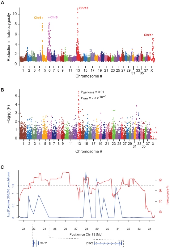
In order to find candidate loci for the breed-specific phenotype (hyaluronanosis), known to be under selective pressure, we screened the genome for signatures of selective sweeps. These sweeps can be recognized as long chromosomal segments with a low degree of heterozygosity within populations [10]. Using 50,000 single nucleotide polymorphisms (SNPs) distributed throughout the dog genome, the level of heterozygosity in windows of ten consecutive SNPs was compared between a set of Shar-Pei (n = 50, all from the US, Table S1) and the average of 24 other canine breeds (n = 230). On four chromosomes (Cfa 5, 6, 13 and X) the reduction in heterozygosity in Shar-Pei was greater than 4-fold the average of control breeds (Figure 2A). The strongest signal of reduced heterozygosity appeared within a 3.7 Mb stretch on chromosome 13 (CanFam 2.0 Chr13 : 23,487,992–27,227,623) (http://genome.ucsc.edu/) near the HAS2 gene, where almost complete homozygosity was observed in Shar Pei (Figure 2C). Here the reduction in heterozygosity was greater than 10-fold in Shar-Pei and several smaller regions showed complete homozygosity. The same region was confirmed to show high levels of homozygosity when the analysis was repeated in 37 additional Shar-Pei dogs sampled from Spain (Table S1) and was overlapping a sweep region reported by others for this breed [11]. The strong signal, together with the known function of HAS2 and its aberrant expression pattern in Shar-Pei, made this region an obvious candidate for the mutation causing the wrinkled skin phenotype (hyaluronanosis).
Fig. 2. The association with Shar-Pei Fever susceptibility and the strongest selective sweep signal co-localize on chromosome 13. 
(A) A 10-fold reduction of heterozygosity was identified on chromosome 13 when comparing Shar-Pei (n = 50) to 24 other canine breeds (n = 230). 50,000 SNPs were used to screen the whole genome using a sliding window approach (see Materials and Methods). (B) A case-control genome-wide association analysis identified a strong peak with several SNPs on chromosome 13 to be in association with Familial Shar-Pei Fever (FSF). After correcting for stratification and multiple testing (100,000 permutations), four SNPs retained significant association (p<0.05; strongest SNP association, CanFam 2.0 chr13: 27,913,803 Mb). Shar-Pei dogs used in the study were strictly classified into groups of affected (n = 22) and unaffected (n = 17) by FSF. (C) SNPs associated with FSF (blue line) are interspersed with the signals of selection (red line). The 39 Shar-Pei and 17,227 SNP common to both analyses were used to generate this graph. In parallel, we performed a genome-wide association study to map the susceptibility locus for FSF, using Shar-Pei strictly classified as FSF affected (n = 24, classification code FSF+A and FSF+, described in Materials and Methods) and unaffected (n = 17, classification code H+, described in Materials and Methods). Five SNPs were significantly associated (best SNP praw = 7.0×10⁻⁷, pgenome = 0.005 based on 100,000 permutations; software package PLINK http://pngu.mgh.harvard.edu/~purcell/plink [12]), all on chromosome 13 (CanFam 2.0 Chr13 : 22.4–30.7 Mb, Figure 2B). After correcting for putative stratification, two outlier cases were removed (Figure S1) and the same SNPs, forming the same signal of association remained (best SNP praw = 2.3×10⁻⁶, pgenome = 0.01; Table S2) with a genomic inflation factor of 1.2. When the association signal and the sweep signal were compared they appeared interspersed, so that individual SNPs were either part of homozygous regions or showed association with FSF (Figure 2C). It was therefore difficult to determine exactly where the strongest association fell, as variation is required to detect association.
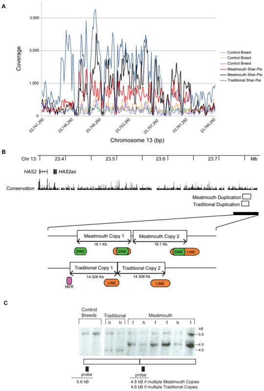
Targeted sequence capture technology was used to further investigate the sweep signal and to search for the hyaluronanosis causative mutation. We resequenced 1.5 Mb around and upstream of our candidate gene, HAS2 (CanFam 2.0 Chr13 : 22,937,592–24,414,650) in four Shar-Pei (two meatmouth type with high serum HA levels and two traditional type) and three control dogs from other breeds. The obtained sequences were mapped to the boxer reference sequence providing at least 5X coverage for 96–98% of the resequenced region in each individual. The targeted region also included the large intergenic noncoding RNA, HAS2 antisense (HAS2as; Table S3) which has been proposed as a negative post-transcriptional regulator of HAS2 mRNA [13]. After masking repetitive sequences we identified ∼670 indels and ∼1,500 SNP in each dog (Table S4) as well as two overlapping duplications in the Shar-Pei (Figure 3A). Nine mutations (eight SNPs and one indel) located in conserved elements as well as two SNPs possibly regulating transcription, were selected for further investigation due to their unique pattern in the sequenced Shar-Pei dogs. Additional genotyping in Shar-Pei and dogs from other breeds (Tables S1, S5) showed these mutations were not specific to Shar-Pei and the variants were subsequently excluded as causative.
Fig. 3. The identification of two breed-specific duplications in Shar-Pei. 
(A) Targeted resequencing of a 1.5 Mb region on chromosome 13 identified a duplication with on average 3.5–4.5X higher read coverage in two meatmouth Shar-Pei (black and red), compared to three control breeds (green, Standard Poodle; orange, Neapolitan Mastiff and purple, Pug). A shorter duplication was detected in the traditional Shar-Pei (blue). (B) The meatmouth duplication was determined to be 16.1 Kb long (CanFam 2.0 Chr13: 23,746,089–23,762,189) with both breakpoints located in repeats (a SINE and a LINE) and with an insertion of 7 bp separating different copies. The duplication in the traditional Shar-Pei overlapped the meatmouth duplication and was slightly shorter, 14.3 Kb long (CanFam 2.0 Chr13: 23,743,906–23,758,214). In this case the copies were separated by 1 bp but were still anchored in repeat motifs (c) Southern blot analysis using BsrGI digested gDNA from Shar-Pei and control breeds confirmed the existence of two duplication types in Shar-Pei. One meatmouth dog (lane 6) contained both duplication types. Individuals were classified as healthy (h) or as affected by Familial Shar-Pei Fever (f). The two duplications were named after the Shar-Pei type in which they were first identified. The “meatmouth” duplication was the larger fragment, 16.1 Kb (CanFam 2.0 Chr13 : 23,746,089–23,762,189) with breakpoints located in repeats (a SINE at the centromeric end and a LINE at the telomeric end) and individual copies separated by seven base pairs (Figure 3B). The “traditional” duplication was 14.3 Kb (CanFam 2.0 Chr13 : 23,743,906–23,758,214) and was identified in the two Shar-Pei with a less accentuated skin phenotype (Figure 3B). We first examined the duplications via Southern blot with control breeds (n = 2), traditional (n = 2) and meatmouth Shar-Pei (n = 6) (Figure 3C). As the digest cut outside and within both duplications, we were able to observe the absence of the variants from control breeds and separate restriction patterns in traditional and meatmouth type Shar-Pei. Interestingly, one meatmouth dog contained both duplication types (Figure 3C lane 6 and confirmed by PCR across break points, data not shown). Two copy number assays were developed to quantify these elements. The first (CNV-E) measured only the meatmouth duplication whilst the second (CNV-748), detected both the traditional and meatmouth duplications. Copy number analysis was estimated as the relative fold enrichment (ΔΔCt) between an amplicon within the duplication and one outside the duplication in a housekeeping gene. Assay CNV-E was run on 90 Shar-Pei and 73 dogs from 24 other breeds (Table S1) and assay CNV-748 on a subset of 44 Shar-Pei and 14 dogs from other breeds. Assay CNV-748 demonstrated that both the traditional and meatmouth duplications are unique to the Shar-Pei breed (Figure 4 and Figure S2).
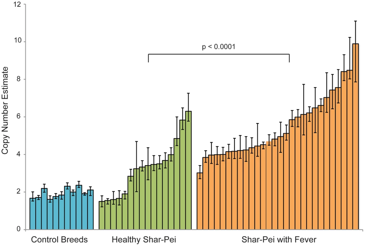
Fig. 4. The relationship between copy number estimate and susceptibility to Familial Shar-Pei Fever. 
A significant correlation (p = <0.0001, Mann Whitney test) was seen when the meatmouth copy number in unaffected Shar-Pei (n = 16, H+) and individuals affected by FSF (n = 28, FSF+ and FSF+ A) were compared. Based on this limited sample size, most dogs with more than six copies had fever whereas most dogs with less than four copies did not. We used the results of both assays to search for a relationship between Familial Shar-Pei Fever (FSF) and either meatmouth copy number (Assay CNV-E), traditional copy number (the normalized difference between CNV-748 and CNV-E) or total traditional+meatmouth copy number (Assay CNV-748). Shar-Pei dogs were strictly classified as affected by FSF (n = 28, FSF+A and FSF+) or unaffected by FSF (n = 16, H+). The most significant association was found when only the meatmouth copy number was considered (p<0.0001, Figure 4) although a weaker association with total copy number (p<0.01) was also seen. The observed association between fever and meatmouth copy number, despite the very high homozygosity in this region, strongly suggests that a high copy number is not just a genetic marker for FSF but is causally related to the development of disease.
Of the 153 dogs analyzed with the meatmouth copy number assay, 31 Shar-Pei and 18 control animals also had serum measures of HA available. No clear association was detected between HA levels and copy number (Figure S3), however the mean HA level in Shar-Pei with ≥ six copies was 905±403 ug/L (n = 21), whilst Shar-Pei with fewer copies had a mean concentration of 770±494 ug/L (n = 12) and control breeds had HA serum levels of 206±145 ug/L (n = 19). Interestingly, the three traditional Shar-Pei dogs had serum HA levels between 73 and 266 ug/L, which fell within the normal range [4].
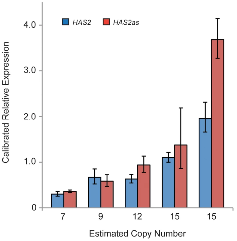
The link between copy number and the expression of HAS2 and HAS2as was examined on a smaller scale using dermal fibroblasts cultured from six separate meatmouth Shar-Pei. The expression of both genes was calibrated against the Shar-Pei with lowest copy number (CNV estimate = 5) and both genes showed an increasing trend of expression with copy number (Figure 5). These data suggest that a regulatory element for HAS2 is located in the duplicated region, however the interpretation of the HAS2as result is less clear. A single study of a human osteosarcoma cell line demonstrated that the expression of two isoforms of HAS2as were able to reduce HAS2 expression, and so these mRNAs may act as regulators of HA production [13]. Our data could indicate that HAS2as expression is also influenced by a regulator element in the duplication, or that HAS2as is up-regulated in response to HAS2 levels. If either of these scenarios were true, it is possible that if RNA expression were measured at multiple time points we would see temporal HAS2 repression. It could also be that the interaction between canine fibroblast HAS2 and HAS2as does not mirror the human system and that the canine antisense mRNA is non-functional. At present our results must be considered as preliminary and it is clear that further exploration of the interaction between canine HAS2 and HAS2as is required.
Fig. 5. Expression analysis reveals a trend of increased HAS2 and HAS2as expression with copy number. 
Expression levels were measured in dermal fibroblasts that were cultured from individual Shar-Pei skin biopsies. The individual with the lowest copy number (CNV = 5) was used to calibrate each assay. Discussion
Here we have identified a 16.1 Kb duplication located approximately 350 Kb upstream of HAS2. This is clearly a derived mutation since it occurs as a single copy sequence in other dog breeds. We postulate that this is a causative mutation associated with both hyaluronanosis and Shar-Pei fever, as the observed correlation between copy number and susceptibility to Shar-Pei fever was not expected if this was a linked, neutral polymorphism. We suggest that the unique region of the meatmouth type duplication identified in Shar-Pei contains one or more regulatory elements that alter the expression of HAS2. It appears possible that as the duplication copy number increases, so does the copy number of potential enhancer elements within the duplication, likely leading to a higher expression of HAS2 and elevated HA levels, and resulting in the development of hyaluronanosis in this breed. We propose a scenario whereby the traditional duplication arose de novo in the traditional type of Shar-Pei causing a milder skin phenotype. This event made the region unstable and allowed the second meatmouth duplication to occur. Breeders subsequently selected the meatmouth duplication as a higher copy number enhanced the phenotypic effect in appearance. However, it is not yet possible to say whether the meatmouth duplication first occurred at low frequency in the Chinese Shar-Pei population and quickly rose during breeding in America, or if the mutation occurred spontaneously during breed expansion in the West.
Tandem duplications are notoriously unstable and may show copy number variation due to unequal crossing-over, as is clearly illustrated by the copy number variation of a 450 Kb duplication associated with dominant white colour in pigs [14]. The meatmouth Shar-Pei duplication adds to the list of copy number variants (CNVs), which affect phenotypic traits in domestic animals (e.g. dominant white in pigs [14], gray color in horses [15], the hair-ridge in Rhodesian ridgeback dogs [16], and pea-comb in chicken [17]), several of which are linked not only to the desirable trait but also to disease. Interestingly, all of these except pea-comb, represent novel duplications derived from single copy sequences. This is in contrast to most reported CNVs in humans, which are mainly benign and represent expansions or contractions of duplicated sequences [18].
Although we failed to find a significant correlation between serum HA levels and copy number, this does not exclude our proposed hyaluronanosis scenario. Difficulties in correlating fluctuating serum levels of HA with other clinical and biomedical parameters have also been reported in many human studies, where no or only weak correlations were observed [19], [20]. We have shown that the 16.1 Kb duplication appears only in meatmouth Shar-Pei, a breed type that has elevated levels of HA compared to both traditional Shar-Pei and other breeds, and that copy number correlates with a breed-specific syndrome associated with excessive HA deposition and the over expression of a HA synthesizing gene. Because HA is primarily a component of the extracellular matrix, serum measurements may only broadly reflect total body HA.
Hyaluronan can bind to several cellular receptors (e.g. CD44, RHAMM and layilin), however it is the interaction between CD44 and HA which acts as a biological regulator, differentially modulating the cellular microenvironment in response to homeostatic versus inflammatory conditions [21]. Alterations in the balance between native high molecular weight HA versus fragmented HA may result in activation of innate immunity. HA has been linked to sterile inflammation as an endogenous response molecule to sterile tissue injury [21]. Shorter fragments of HA can be generated by environmental insults such as sterile trauma [22], reactive oxidative species (ROS) [23], or pathogenic hyaluronidases, and it is these low molecular weight fractions which can become pro-inflammatory danger associated molecular pattern (DAMP) molecules [22], [24] mimicking microbial surface molecules.
Using a mouse model, Yamasaki and colleagues [25] showed that HA can interact with the cell through two separate pathways that culminate in the release of IL-1β, which together with IL-6, is one of the main promoters of fever. In the first route, CD44 bound HA is degraded at the plasma membrane by hyaluronidase-2 (HYAL2) prior to endocytosis and further cleavage by lysosomal hyaluronidase-1 (HYAL1). The resultant small intracellular oligosaccharides of HA activate the NLRP3 inflammasome, a multiprotein complex consisting of the NLRP3 scaffold, the ASC adaptor and caspase-1 [26]. In the second arm, the CD44-HA complex activates toll like receptors 2 and 4 (TLR2 and 4), leading to intracellular IL-1β mRNA transcription and the formation of pro-IL-1β. Activation of the NLRP3 inflammasome by HA oligosaccharides allows cleavage of this pro-IL-1β by caspase-1 and subsequent release of IL-1β. The NLRP3 inflammasome is present in the cytosol of many cells including monocytes, macrophages and mast cells, and has been implicated in the pathogenesis of numerous autoinflammatory diseases in humans including the cryopyrin-associated periodic syndromes which result from mutations in NLRP3/CIAS1 [26].
The actual role of excessive HA in Shar-Pei needs to be investigated further. Shar-Pei may experience exogenous fragmentation of their over-abundant HA from sterile or pathogenic trauma. This, plus endogenous degradation of excessive native HA, may contribute to induction of recurrent episodes of fever and inflammation. Acute fever events in Shar-Pei respond rapidly to dipyrone, a potent antipyretic and analgesic pyrazolone, which has been demonstrated to inhibit IL-1β induced fever [27]–[29 and Linda Tintle unpublished data]. It is therefore not surprising that the strong selection on the hyaluronanosis phenotype, with increased levels of cutaneous HA, may predispose Shar-Pei to autoinflammation, potentially contributing to other pathologies seen in this breed. One such example is renal medullary amyloidosis. Histopathologically, kidneys of Shar-Pei in renal failure have multifocal non-suppurative tubulointerstitial nephritis with fibrosis. Medullary amyloidosis predominates and glomerular deposition, although consistent, is highly variable in its extent [8], [30]. The renal medulla is naturally HA rich and enhanced renal interstitial HA accumulation can be coupled to inflammatory responses, such as ischemia-reperfusion injury, transplant-rejection, tubulointerstitial inflammation and diabetes [31]. In addition, Shar-Pei are prone to mast cell disease including mast cell tumors [32], [33]. The binding of HA to CD44 has been shown to play a critical role in regulation of murine cutaneous and connective tissue mast cell proliferation [34]. As the CD44-HA interaction may modulate local immune responses through regulation of mast cell functions [35], excessive HA and its subsequent damage and degradation may play a role also in the Shar-Pei breed’s predilection for allergic skin disease and other mast cell driven inflammation.
This study suggests that HAS2 dysregulation can trigger a periodic fever syndrome in dogs and therefore it will be relevant to examine the approximately 60% of human fever patients who currently have unexplained disease. Previously, the role of hyaluronan in sterile inflammation has focused on HA signaling and degradation; for example a deficiency of hyaluronidase causing mucopolysaccharidosis type IX in humans has some autoinflammatory features [36]. However by directly implicating HAS2 in inflammation, we suggest that a reexamination of genes further up the biosynthetic pathway, such as those involved in HA synthesis and polymerization is called for. In addition, the canine mutation appears regulatory in nature and therefore regulators of HA should be also be included in a broader scope pathway analysis of human patients with unexplained autoinflammatory disease.
Finally, this study illustrates how copy number variations can shape phenotypic traits and how strong artificial selection for certain phenotypic traits may not only affect the desired trait but also the health of the animal.
Materials and Methods
Samples and diagnostic procedure
All dog samples were collected from pet dogs after owner consent following the ethical approval protocols (SLU, Dnr: C103/10, MIT 0910-074-13). DNA was extracted from blood samples using QIAamp DNA Blood Midi Kit (QIAGEN) or PureLink Genomic DNA kit (Invitrogen). All dogs, their breed type, geographic origin, health status and experiment in which they were utilized are listed in Table S1.
Classification of Shar-Pei fever: Purebred Shar-Pei individuals were divided into the following six groups based on their medical records and evidence by owner and/or veterinarian:
1. FSF+A, the individual had experienced recurrent episodes of high fever accompanied by inflammation of joints from an early age (less than one year old). Additionally, post-mortem examination detected depositions of amyloid in kidneys and/or liver (amyloidosis).
2. FSF+, the individual had experienced recurrent episodes of high fever accompanied with inflammation of joints from an early age (less than one year old).
3. Atypical FSF, the individual had experienced occasional unexplained fever episodes or recurrent episodes with a late onset (greater than three years old).
4. H+, the individual had never experienced unexplained fever and/or inflammation, was older than five years old at the time of sampling and also lacked first-degree relatives that could be classified into the groups FSF+A, FSF+ or Atypical FSF.
5. H-, the individual had never experienced unexplained fever and/or inflammation but was younger than 5 years at the time of sampling and/or had first-degree relatives that could be classified into the groups FSF+A, FSF+ or Atypical FSF.
6. Unknown, the individual’s medical record was not available.
Hyaluronanosis: Serum Hyaluronic Acid (HA) concentration was used as a proxy for hyaluronanosis but no distinct cut-off value was established. However, dogs with normal and abnormal concentrations of serum HA were interpreted as before [4]. HA measurements were performed using the Hyaluronan ELISA kit (Echelon Biosciences INC) according to the manufacturer’s instructions. The absorbance was read at 405 nm, and a semi-log standard curve was used to calculate hyaluronic acid concentrations.
Homozygosity and genome-wide association mapping
A whole genome scan was performed with two array types, the 27K (v1) and 50K (v2) canine Affymetrix SNP chips. Results were called using Affymetrix’s snp5-geno-qc software. The 50K array was used when the rate of heterozygosity was calculated for US Shar-Pei separately and for a reference group of 24 other breeds. The ratio of heterozygosity in 10 SNP (≈1 Mb) sliding windows between the two groups was used as a measure of relative heterozygosity. To look for regions of homozygosity within the Shar-Pei genome only, the software package PLINK [12] was used. This was performed both for the 50 K array with 50 US Shar-Pei and replicated for 37 Spanish Shar-Pei using 22,362 SNPs genotyped with the Illumina CanineSNP20 BeadChip. These data were collected with an Illumina BeadStation scanner and genotypes were scored using GenomeStudio. Regions of homozygosity were defined if shared across all Shar-Pei samples.
A case-control association analysis using 17,227 SNP common to both the 27K and 50K arrays (MAF>0.05, call rate >75%) was performed in Shar-Pei classified as affected (FSF+A and FSF+, n = 39) or unaffected (H+, n = 17) by Shar-Pei fever. The software package PLINK [12] was used for the analyses and to ensure genome-wide significance, p-values were corrected for multiple testing. Values used are the max (T) empirical p-values obtained after 100,000 permutations. To assess whether signals from the two genome scans overlapped, the 39 Shar-Pei with unambiguous phenotypes were analyzed with the 17,227 SNPs common to both SNP platforms.
Targeted resequencing
Targeted capture of the 1.5 Mb candidate region (CanFam 2.0 Chr13 : 22,937,592–24,414,650) was performed using a 385K custom-designed sequence capture array from Roche NimbleGen. Hybridization library preparation was performed as following: Genomic DNA (15–20 µg) was fragmented using sonication; blunting of DNA fragments using T4 DNA Polymerase, Klenow Fragment and T4 Polynucleotide Kinase; adding A-overhangs using Klenow Fragment exo− and ligation of adaptors using T4 DNA Ligase with Single-read Genomic Adapter Oligo Mix (Illumina). All enzymes were purchased from Fermentas and used following manufacturers instructions. Purification steps were performed using QIAquick PCR Purification Kit (QIAGEN). Hybridization was performed following the manufacturer’s instructions without amplification of the fragment library prior to hybridization. Eluted captured DNA and uncaptured libraries were amplified using Phusion High Fidelity PCR Master Mix (Finnzymes) and the SYBR Green PCR Master Mix (Applied Biosystems) was used to estimate the relative fold-enrichment. Capture libraries with the estimated enrichment-factor of >200 were sequenced using Genome Analyzer (Illumina) and obtained sequences were aligned to CanFam 2.0 [37] and to the targeted region using Maq assembly (http://maq.sourceforge.net/) [38]. For each individual, sequence coverage was calibrated by dividing the coverage in 100 bp windows by the average coverage for the total region. Three control breeds (Pug, Neapolitan Mastiff, Standard Poodle) and two of each type of Shar-Pei (meatmouth type and traditional type) were sequenced. The two traditional type Shar-Pei were sequenced at different read lengths but were aligned using the same strict criteria (allowing two mismatches per read) and therefore vary in the percentage of mapped reads as well as coverage when compared to the other individuals. Individual 7 (Table S4) was sequenced from whole genome amplified material and this may have impacted the ability to map reads and detect SNPs. This individual was not plotted in Figure 3A, but was used in downstream analyses.
Polymerase Chain Reaction (PCR) and Sanger Sequencing
All primers used were designed using Primer3 (http://frodo.wi.mit.edu/primer3/) [39] and are listed in Table S6. PCR and Sanger Sequencing was performed to investigate putative mutations (ten SNPs and one indel) and were carried out with 20 ng genomic DNA using AmpliTaq Gold DNA Polymerase (Applied Biosystems) following the manufacturer’s instructions. The amplification of the copy number variant (CNV) breakpoints was performed with 400 ng of DNA and a Long-range PCR with Expand Long Template PCR System Mix 1 (Roche), cloned using Zero Blunt TOPO Cloning Kit (Invitrogen) and plasmid DNA prepared using QIAprep Spin Miniprep Kit (QIAGEN). PCR products and plasmids were sequenced using capillary electrophoresis 3730xl (Applied Biosystems), aligned and analyzed using CodonCode Aligner version 2.0.6 (CodonCode).
Southern blot analysis
Four micrograms of genomic DNA from each sample was digested with BsrGI (New England BioLabs) and separated on a 0.7% agarose gel. A 910 bp probe (targeting CanFam 2.0 Chr13 : 23,746,12–23,747,522) was used to detect the duplicated region.
Copy number assay
Estimation of copy number was performed using the comparative CT (ΔΔCT) relative quantification method and a calibrator animal (German Shepherd 95). The duplex reaction contained a primer limited copy number assay (CNV-E: 300 nM each of forward and reverse primers, 250 nM FAM labeled MGB probe; CNV-748 : 50 nM of forward and 300 nM reverse primers, 250 nM FAM labeled MGB probe, Applied Biosystems) and a reference assay designed to C7orf28B (900 nM of forward and reverse primers, 250 nM VIC and TAMRA labeled probe, Applied Biosystems). Real Time PCR was performed in quadruplet using 10 ng of gDNA, Genotyping Master Mix (Applied Biosystems) and a 7900 HT Real Time PCR machine (Applied Biosystems). The PCR primers used and dogs evaluated can be found in Tables S4 and S1 respectively.
Fibroblast cultures
Cultures of dermal fibroblasts were established from skin samples of Shar-Pei dogs as described previously [40]. Skin samples were well shaved and cleaned with 70% EtOH/Betadine before biopsy and cell isolation. Fat tissue and blood vessels were removed from the skin and then samples were washed with PBS, cut into small fragments (0.5 cm2) and digested with dispase II solution (Boehringer Mannheim) for 16 h at 4°C. The next day, after incubation for 30 min at 37°C in the same solution, the dermis was separated from the epidermis. Washed dermal samples were chopped into 1 mm3 fragments and incubated for 140 min in 15 ml of DMEM per gram of skin containing 30 mg bacterial collagenase (Gibco), 18 mg hyaluronidase, 12 mg pronase, 1.5 mg DNAse, supplemented with bovine albumin (all from Sigma) and antibiotics. After digestion, cutaneous cells were washed with PBS and grown in a humidified atmosphere at 37°C with 5% CO2 for two days. Medium was changed twice a week and cells were used at passages two-five.
Gene expression analysis
RNA extraction from fibroblast cultures was performed as described elsewhere [41]. 500 ng of RNA was reverse transcribed using the High-Capacity cDNA Archive Kit (Applied Biosystems) with random primers and following the manufacturer’s instructions. Two assays were designed to target HAS2 and HAS2as cDNA, respectively. Real Time PCR in a volume of 20 ul was performed in duplicate using SYBR Green PCR Master Mix (Applied Biosystems) and primers at 300 nM in a 7900 HT Real-Time PCR system (Applied Biosystems) with standard cycling. PCR specificity assessment was performed by adding a dissociation curve analysis at the end of the run. Each amplification run contained negative controls. Relative fold-enrichment was performed using the comparative ΔCT-method with Glucose-6-phosphate dehydrogenase (G6PD) for normalization.
Web resources
http://pngu.mgh.harvard.edu/~purcell/plink/
http://frodo.wi.mit.edu/primer3/
Supporting Information
Zdroje
1. FraserJR
LaurentTC
LaurentUB
1997 Hyaluronan: its nature, distribution, functions and turnover. Journal of Internal Medicine 242 27 33
2. Wheeler-JonesCP
FarrarCE
PitsillidesAA
2010 Targeting hyaluronan of the endothelial glycocalyx for therapeutic intervention. Current Opinion in Investigational Drugs 11 9 997 1006
3. LaurentTC
FraserJRE
1992 Hyaluronan. FASEB J 6 2397 2404
4. ZannaG
FondevilaD
BardagiM
DocampoMJ
BassolsA
2008 Cutaneous mucinosis in shar-pei dogs is due to hyaluronic acid deposition and is associated with high levels of hyaluronic acid in serum. Vet Dermatol 19 314 318
5. RamsdenCA
BankierA
BrownTJ
CowenPSJ
FrostGI
2000 A new disorder of hyaluronan metabolism associated with generalized folding and thickening of the skin. J of Ped 36 62 68
6. WeigelPH
HascallVC
TammiM
1997 Hyaluronan synthases. J Biol Chem 272 13997 4000
7. ZannaG
DocampoMJ
FondevilaD
BardagiM
BassolsA
2009 Hereditary cutaneous mucinosis in Shar-Pei dogs is associated with increases hyaluronan synthase-2 mRNA transcription by cultured dermal fibroblasts. Vet Dermatol 20 377 382
8. RivasAL
TintleL
KimballES
ScarlettJ
QuimbyFW
1992 A canine febrile disorder associated with elevated interleukin-6. Clin Immunol Immunopathol 64 36 45
9. StojanovS
KastnerDL
2005 Familial autoinflammatory diseases: genetics, pathogenesis and treatment. Curr Opin Rheumatol 17 5 586 599
10. SmithJM
HaighJ
1974 The hitchhiking effect of a favorable gene. Genetic Research 23 23 35
11. AkeyJM
RuheAL
AkeyDT
WongAK
ConellyCF
2010 Tracking footprints of artificial selection in the dog genome. PNAS 19 1160 1165
12. PurcellS
NealeB
Todd-BrownK
ThomasL
FerreiraMA
2007 PLINK: a tool set for whole-genome association and population-based linkage analyses. Am J Hum Genet 81 559 575
13. ChaoH
SpicerAP
2005 Natural Antisense mRNAs to Hyaluronan Synthase 2 Inhibit Hyaluronan Biosynthesis and Cell Proliferation. J Biol Chem 19 27513 27522
14. GiuffraE
TörnstenA
MarklundS
Bongcam-RudloffE
ChardonP
2002 A large duplication associated with dominant white color in pigs originated by homologous recombination between LINE elements flanking KIT. Mamm Genome 13 10 569 77
15. Rosengren PielbergG
GolovkoA
SundströmE
CurikI
LennartssonJ
2008 A cis-acting regulatory mutation causes premature hair graying and susceptibility to melanoma in the horse. Nat Genet 40 8 1104 1009
16. Salmon HillbertzNHC
IsakssonM
KarlssonEK
HellménE
PielbergGR
2007 A duplication of FGF3, FGF4, FGF9 and ORAOV1 causes the hair ridge and predisposes to dermoid sinus in Ridgeback dogs. Nat Genet 38 11 1318 1320
17. WrightD
BoijeH
MeadowsJR
Bed'homB
VieaudA
2009 Copy number variation in intron 1 of SOX5 causes the pea-comb in chickens. PLoS Genet 5 6 1000512 doi:10.1371/journal.pgen.1000512
18. SebatJ
LakshmiB
MalhotraD
TrogeJ
Lese-MartinC
2007 Strong association of de novo copy number mutations with autism. Science 316 445 449
19. GoldbergRL
HuffJP
LenzME
GlickmanP
KatzR
1991 Elevated plasma levels of hyaluronan in patients with osteoarthritis and rheumatoid arthritis. Arthritis Rheum 34 799 807
20. HedinPJ
WeitoftT
HedinH
Engström-LaurentA
SaxneT
1991 Serum concentration of hyaluronan and proteoglycans in joint disease. Lack of association. J Rheumatol 18 1601 1605
21. PuréE
AssoianRK
2009 Rheostatic signaling by CD44 and hyaluronan. Cell Signal 21 651 655
22. HascallVC
MajorsAK
De La MotteCA
EvankoSP
WangA
2004 Intracellular hyaluronan: a new frontier for inflammation? Biochim Biophys Acta 1673 3 12
23. SternR
Asari AA
SugaharaKN
2006 Hyaluronan fragments: An information-rich system. Eur J Cell Biol 85 8 699 715
24. EberleinM
SchneiberKA
BlackKE
CollinsSL
Chan-LiY
2008 Anti-oxidant inhibition of hyaluronan fragment-induced inflammatory gene expression. J Inflamm 5 20
25. YamasakiK
MutoJ
TaylorKR
CogenAL
AudishD
2009 NLRP3/Cryopyrin is necessary for Interleukin-1β (IL-β) release in response to hyaluronan, an endogenous trigger in inflammation in response to injury. J Biol Chem 284 19 12762 12771
26. KastnerDL
AksentijevichI
Goldbach-ManskyR
2010 Autoinflammatory Disease reloaded: A clinical perspective. Cell 140 784 790
27. ShimadaSG
OtternessIG
StittJT
1994 A study of the mechanism of action of the mild analgesic dipyrone. Agents Actions 41 188 192
28. deSouzaGE
CardosoRA
MeloMC
FabricioAS
SilvaVM
2002 A comparative study of the antipyretic effects of indomethacin and dipyrone in rats. Inflamm Res 51 1 24 32
29. PradaJ
DazaR
ChumbersO
LoayzaI
HuichoL
2006 Antipyretic efficacy and tolerability of oral ibuprofen, oral dipyrone and intramuscular dipyrone in children: a randomized controlled trial. Sao Paolo Med J 124 3 135 140
30. DiBartolaSP
TarrMJ
WebbDM
GigerU
1990 Familial renal amyloidosis in Chinese Shar Pei dogs. J Am Vet Assos 15 483 487
31. StridhS
KerjaschkiD
ChenY
RugenheimerL
ÅstrandABM
2010 Angiotensin converting enzyme inhibition blocks interstitial hyaluronan dissipation in the neonatal rat kidney via hyaluronan synthase 2 and hyaluronidase 1. Matrix Biol In press
32. LópezA
SpracklinD
McConkeyS
HannaP
1999 Cutaneous mucinosis and mastocytosis in shar-pei. Can Vet J 40 881 883
33. MillerDM
1995 The occurrence of mast cell tumors in young shar-peis. J Vet Diagn Invest 7 360 363
34. TakanoH
NakazawaS
ShirataN
TambaS
FurutaK
2009 Involvement of CD44 in mast cell proliferation during terminal differentiation. Lab Invest 89 4 446 55
35. TanakaS
2010 Targeting CD44 in mast cell regulation. Expert Opin Ther Targets 14 1 31 43
36. Triggs-RaineB
SaloTJ
ZhangH
WicklowBA
NatowiczMR
1999 Mutations in HYAL1, a member of a tandemly distributed multigene family encoding disparate hyaluronidase activities, cause a newly described lysosomal disorder, mucopolysaccharidosis IX. Proc Natl Sci USA 11 6296 6300
37. Lindblad-TohK
WadeCM
MikkelsenTS
KarlssonEK
JaffeDB
2005 Genome sequence, comparative analysis and haplotype structure of the domestic dog. Nature 438 803 819
38. HengL
RuanJ
DurbinR
2008 Mapping short DNA sequencing reads and calling variants using mapping quality scores. Genome Res18 1851 1858
39. RozenS
SkaletskyHO
2000 Primer3 on the WWW for general users and for biologist programmers. Bioinformatics Methods and Protocols: Methods in Molecular Biology Totowa, New Jersey, USA Humana Press 365 386
40. SerraM
BrazísP
PuigdemontA
FondevillaD
RomanoV
2007 Development and characterization of a canine skin equivalent. Exp Dermatol 16 135 142
41. ChomczynskiP
MackeyK
1995 Short technical report. Modification of the TRIZOL reagent procedure for isolation of RNA from Polysaccharide-and proteoglycan-rich sources. Biotechniques 19 6 942 5
Štítky
Genetika Reprodukční medicína
Článek Genetic Regulation by NLA and MicroRNA827 for Maintaining Nitrate-Dependent Phosphate Homeostasis inČlánek c-di-GMP Turn-Over in Is Controlled by a Plethora of Diguanylate Cyclases and PhosphodiesterasesČlánek Viral Genome Segmentation Can Result from a Trade-Off between Genetic Content and Particle Stability
Článek vyšel v časopisePLOS Genetics
Nejčtenější tento týden
2011 Číslo 3
-
Všechny články tohoto čísla
- Whole-Exome Re-Sequencing in a Family Quartet Identifies Mutations As the Cause of a Novel Skeletal Dysplasia
- Origin-Dependent Inverted-Repeat Amplification: A Replication-Based Model for Generating Palindromic Amplicons
- Testing for an Unusual Distribution of Rare Variants
- Limited dCTP Availability Accounts for Mitochondrial DNA Depletion in Mitochondrial Neurogastrointestinal Encephalomyopathy (MNGIE)
- FUS Transgenic Rats Develop the Phenotypes of Amyotrophic Lateral Sclerosis and Frontotemporal Lobar Degeneration
- Repeat Associated Non-ATG Translation Initiation: One DNA, Two Transcripts, Seven Reading Frames, Potentially Nine Toxic Entities!
- Initial Mutations Direct Alternative Pathways of Protein Evolution
- Dopamine Signalling in Mushroom Bodies Regulates Temperature-Preference Behaviour in
- Sensing of Replication Stress and Mec1 Activation Act through Two Independent Pathways Involving the 9-1-1 Complex and DNA Polymerase ε
- Genetic Regulation by NLA and MicroRNA827 for Maintaining Nitrate-Dependent Phosphate Homeostasis in
- Identification of a Novel Type of Spacer Element Required for Imprinting in Fission Yeast
- Chiasmata Promote Monopolar Attachment of Sister Chromatids and Their Co-Segregation toward the Proper Pole during Meiosis I
- Global Analysis of the Relationship between JIL-1 Kinase and Transcription
- H3K9me2/3 Binding of the MBT Domain Protein LIN-61 Is Essential for Vulva Development
- REVEILLE8 and PSEUDO-REPONSE REGULATOR5 Form a Negative Feedback Loop within the Arabidopsis Circadian Clock
- A Novel Unstable Duplication Upstream of Predisposes to a Breed-Defining Skin Phenotype and a Periodic Fever Syndrome in Chinese Shar-Pei Dogs
- Polycomb Repressive Complex 2 Controls the Embryo-to-Seedling Phase Transition
- A Role for Set1/MLL-Related Components in Epigenetic Regulation of the Germ Line
- Genome-Wide Association Analysis Identifies Variants Associated with Nonalcoholic Fatty Liver Disease That Have Distinct Effects on Metabolic Traits
- A Genome-Wide Association Study of Upper Aerodigestive Tract Cancers Conducted within the INHANCE Consortium
- Ancestral Mutation in Telomerase Causes Defects in Repeat Addition Processivity and Manifests As Familial Pulmonary Fibrosis
- Ultra-Deep Sequencing of Mouse Mitochondrial DNA: Mutational Patterns and Their Origins
- Phenotype Restricted Genome-Wide Association Study Using a Gene-Centric Approach Identifies Three Low-Risk Neuroblastoma Susceptibility Loci
- The Toll-Like Receptor Gene Family Is Integrated into Human DNA Damage and p53 Networks
- Polycomb Targets Seek Closest Neighbours
- Widespread Hypomethylation Occurs Early and Synergizes with Gene Amplification during Esophageal Carcinogenesis
- c-di-GMP Turn-Over in Is Controlled by a Plethora of Diguanylate Cyclases and Phosphodiesterases
- Estimating Divergence Time and Ancestral Effective Population Size of Bornean and Sumatran Orangutan Subspecies Using a Coalescent Hidden Markov Model
- Rif1 Supports the Function of the CST Complex in Yeast Telomere Capping
- A Tradeoff Drives the Evolution of Reduced Metal Resistance in Natural Populations of Yeast
- Quantifying the Underestimation of Relative Risks from Genome-Wide Association Studies
- Population-Based Resequencing of Experimentally Evolved Populations Reveals the Genetic Basis of Body Size Variation in
- Triplet Repeat–Derived siRNAs Enhance RNA–Mediated Toxicity in a Drosophila Model for Myotonic Dystrophy
- The FUN30 Chromatin Remodeler, Fft3, Protects Centromeric and Subtelomeric Domains from Euchromatin Formation
- Viral Genome Segmentation Can Result from a Trade-Off between Genetic Content and Particle Stability
- Environmental Sex Determination in the Branchiopod Crustacean : Deep Conservation of a Gene in the Sex-Determining Pathway
- Systematic Detection of Polygenic Regulatory Evolution
- The SUMO Isopeptidase Ulp2p Is Required to Prevent Recombination-Induced Chromosome Segregation Lethality following DNA Replication Stress
- Uncoupling Antisense-Mediated Silencing and DNA Methylation in the Imprinted Cluster
- Role of the Drosophila Non-Visual ß-Arrestin Kurtz in Hedgehog Signalling
- Differential Genetic Associations for Systemic Lupus Erythematosus Based on Anti–dsDNA Autoantibody Production
- COMPASS-Like Complexes Mediate Histone H3 Lysine-4 Trimethylation to Control Floral Transition and Plant Development
- H3 Lysine 4 Is Acetylated at Active Gene Promoters and Is Regulated by H3 Lysine 4 Methylation
- Diverse Roles and Interactions of the SWI/SNF Chromatin Remodeling Complex Revealed Using Global Approaches
- A Bow-Tie Genetic Architecture for Morphogenesis Suggested by a Genome-Wide RNAi Screen in
- Roles of () in Oocyte Nuclear Architecture, Gametogenesis, Gonad Tumors, and Genome Stability in Zebrafish
- A Molecular Phylogeny of Living Primates
- Roles of the Espin Actin-Bundling Proteins in the Morphogenesis and Stabilization of Hair Cell Stereocilia Revealed in CBA/CaJ Congenic Jerker Mice
- A Cholinergic-Regulated Circuit Coordinates the Maintenance and Bi-Stable States of a Sensory-Motor Behavior during Male Copulation
- PLOS Genetics
- Archiv čísel
- Aktuální číslo
- Informace o časopisu
Nejčtenější v tomto čísle- Whole-Exome Re-Sequencing in a Family Quartet Identifies Mutations As the Cause of a Novel Skeletal Dysplasia
- Origin-Dependent Inverted-Repeat Amplification: A Replication-Based Model for Generating Palindromic Amplicons
- FUS Transgenic Rats Develop the Phenotypes of Amyotrophic Lateral Sclerosis and Frontotemporal Lobar Degeneration
- Limited dCTP Availability Accounts for Mitochondrial DNA Depletion in Mitochondrial Neurogastrointestinal Encephalomyopathy (MNGIE)
Kurzy
Zvyšte si kvalifikaci online z pohodlí domova
Současné možnosti léčby obezity
nový kurzAutoři: MUDr. Martin Hrubý
Všechny kurzyPřihlášení#ADS_BOTTOM_SCRIPTS#Zapomenuté hesloZadejte e-mailovou adresu, se kterou jste vytvářel(a) účet, budou Vám na ni zaslány informace k nastavení nového hesla.
- Vzdělávání


