-
Články
Reklama
- Vzdělávání
- Časopisy
Top články
Nové číslo
- Témata
Reklama- Videa
- Podcasty
Nové podcasty
Reklama- Kariéra
Doporučené pozice
Reklama- Praxe
ReklamaHost Reticulocytes Provide Metabolic Reservoirs That Can Be Exploited by Malaria Parasites
Malaria, caused by the Apicomplexan parasites Plasmodium spp., is a deadly disease which poses a huge health and economic burden over many populations in the world, mostly in sub-Saharan Africa and Asia. To design new intervention strategies and to improve upon existing drugs against malaria, it is useful to understand the biochemistry of the Plasmodium parasite and its metabolic interplay with the host. Some species of Plasmodium such as P. vivax grow exclusively in reticulocytes (immature erythrocytes) whereas others e.g. P. falciparum will also readily multiply in mature erythrocytes. We asked the questions, do these two classes of host cell offer different resources for parasite survival and could these resources influence antimalarial drug efficacy? We used metabolomics to compare rodent reticulocytes and mature erythrocytes and identified that the metabolome of the former is more diverse and enriched. Gene disruption in the reticulocyte preferring rodent malaria parasite P. berghei was used to demonstrate that Plasmodium can utilise the elements of the metabolic reserves of reticulocytes that mature erythrocytes cannot provide. Our data suggests that the availability of the reticulocyte metabolome might reduce or block the efficacy of antimalarial drugs that target parasite metabolism and drugs tested against P. falciparum might have significantly reduced activity against P. vivax.
Published in the journal: . PLoS Pathog 11(6): e32767. doi:10.1371/journal.ppat.1004882
Category: Research Article
doi: https://doi.org/10.1371/journal.ppat.1004882Summary
Malaria, caused by the Apicomplexan parasites Plasmodium spp., is a deadly disease which poses a huge health and economic burden over many populations in the world, mostly in sub-Saharan Africa and Asia. To design new intervention strategies and to improve upon existing drugs against malaria, it is useful to understand the biochemistry of the Plasmodium parasite and its metabolic interplay with the host. Some species of Plasmodium such as P. vivax grow exclusively in reticulocytes (immature erythrocytes) whereas others e.g. P. falciparum will also readily multiply in mature erythrocytes. We asked the questions, do these two classes of host cell offer different resources for parasite survival and could these resources influence antimalarial drug efficacy? We used metabolomics to compare rodent reticulocytes and mature erythrocytes and identified that the metabolome of the former is more diverse and enriched. Gene disruption in the reticulocyte preferring rodent malaria parasite P. berghei was used to demonstrate that Plasmodium can utilise the elements of the metabolic reserves of reticulocytes that mature erythrocytes cannot provide. Our data suggests that the availability of the reticulocyte metabolome might reduce or block the efficacy of antimalarial drugs that target parasite metabolism and drugs tested against P. falciparum might have significantly reduced activity against P. vivax.
Introduction
The malaria-causing apicomplexan parasites Plasmodium spp. have a dynamic life cycle which is reflected in stage-specific morphologies, transcriptomes, proteomes and metabolomes [1–8]. These changes, particularly in their metabolome, reflect the nutritional needs and biological processes of the parasite during intracellular development that in turn influences, or is influenced by, the physiological state of the host cell [6]. Perhaps due to their parasitic life-style, Plasmodium spp. have a simplified and reduced metabolic capacity when compared to higher non-parasitic organisms. They are auxotrophic for purines, vitamins and many amino acids [9,10], but have retained core pathways of carbon metabolism such as glycolysis [11], the citric acid cycle [7,12], lipid synthesis [13,14], the pentose phosphate pathway [15], pyrimidine biosynthesis [16] and glycosylation [17]. Plasmodium spp. are obligate intracellular parasites and their metabolism is interlinked with that of their host cell and is heavily dependent on the availability of external nutrients. As a result, intracellular Plasmodium establish systems such as the new permeation pathways with the purpose of accessing host cell and environmental nutrients [18]; in fact the parasite genome encodes >120 predicted membrane transport proteins, a subset of which are located on the plasma membrane [19].
Erythrocyte invasion is a prerequisite for establishment of infection by Plasmodium merozoites and the roles of different merozoite and host surface proteins in this invasion process have been intensively studied [20–25]. Multiple partially overlapping erythrocyte invasion pathways have been described in P. falciparum with consequent functional redundancy [26]. Many Plasmodium spp. including P. falciparum preferentially invade reticulocytes [27] which is also capable of invading and replicating within all stages of erythrocyte development including mature cells. However, P. vivax has a strict requirement for growth in reticulocytes, expresses reticulocyte binding proteins [28] and requires a host Duffy blood group glycoprotein for invasion [29]. P. vivax infection causes accelerated remodelling of very young reticulocytes, a process that normally takes 24 hours in uninfected reticulocytes [30]. The rodent model malaria parasite, P. berghei is also 150 times more likely to invade reticulocytes and establish infection in the presence of equal numbers of mature erythrocytes and reticulocytes [31] and has therefore been long thought of as a suitable model for P. vivax blood stage biology [32].
Mature erythrocytes, comprising almost 98% of the circulating red blood cells, can be considered “simplified” cells; they are metabolically active but lack intracellular organelles found in the bone marrow erythroid precursors cells [33] and enucleated reticulocytes (maturing erythrocytes) that are present in peripheral circulation [34]. Reticulocytes undergo many changes after their release into the peripheral circulation as they mature and this is associated with a 20% decrease in total surface area and acquisition of a biconcave shape with consequent increase in shear membrane resistance, the progressive loss of organelles (mitochondria, ribosomes and lysosomes), the loss or reduced abundance of up to 30 membrane proteins, and decreased levels of membrane cholesterol [34,35]. This maturation process is associated with a general streamlining of cellular metabolism; mature erythrocytes are highly dependent on glycolysis [36] and the pentose phosphate pathway [37] for both energy and redox balance and lack many other pathways of carbon metabolism, such as citric acid cycle [38]. Reticulocytes are thus expected to contain a richer repertoire of carbon sources and other essential nutrients than mature erythrocytes which might be exploited or even required by reticulocyte preferent Plasmodium spp.
Limited comparative metabolomics of the erythroid lineage has been attempted before but focussed on sickle cell disease and cord blood reticulocyte physiology [39,40]. Therefore, in order to establish whether there are metabolic differences between reticulocytes and mature erythrocytes that could influence the tropism of different Plasmodium spp., we undertook a non-targeted, high coverage, comprehensive analysis of the metabolomes of these host cells. Comparison of the metabolomes of very young, uninfected rat and human reticulocytes and their mature erythrocyte counterparts revealed major biochemical differences that could be exploited by intracellular parasite stages. This was tested using reverse genetics to disrupt parasite metabolism and establish the broad ability of P. berghei to utilise the products of reticulocyte metabolism and (in part) explain differing profiles of drug susceptibility between parasites in mature erythrocyte and reticulocyte environments.
Results
The reticulocyte metabolome is more complex than that of the mature erythrocyte
Induction of reticulocytosis was achieved through administration of phenylhydrazine-HCl (PHZ, 100 mg/kg body weight) to Wistar rats and cells were harvested when the percentage of reticulocytes in the peripheral blood reached a maximum at day 5 (~35% reticulocytes). This was monitored by FACS analysis using the reticulocyte surface marker transferrin receptor (CD71), which is lost as reticulocytes mature[35]. More than 90% of the 35% reticulocyte population generated by PHZ treatment were CD71-high at the time of harvest (Fig A-A in S1 Text) corresponding to the youngest of the four forms of reticulocytes that have been identified [39] and are from here on referred to as Reticulocyte enriched Erythrocyte Population (REP) Material was also collected for comparison with blood from non-enriched (~1% reticulocytes) animals - wild type Erythrocyte Population (wtEP) (Fig 1A). All samples were uniformly depleted of leucocytes.
Fig. 1. Comparison of Reticulocyte enriched Erythrocyte Population (REP) and wild type Erythrocyte Population (wtEP) reveals metabolite enrichment in rodent and human reticulocytes. 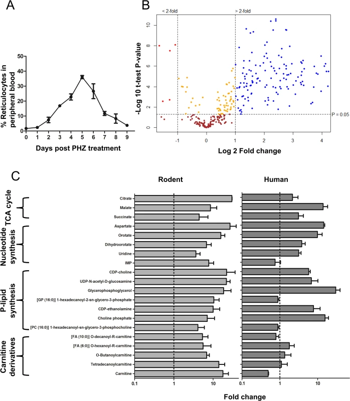
A. Dynamics of reticulocyte enrichment in peripheral blood in vivo followed by Phenylhydrazine-HCl (phz) treatment of mice. Reticulocytes were harvested at day 5 post phz treatment. The error is given as the standard deviation (S.D.) of 3 independent biological replicates. B. Volcano plot showing distribution of putative metabolites according to their fold change in abundance in REP vs wtEP in rodent blood. All significant changes are represented above the broken horizontal line. Coloured dots indicate metabolites which are: Blue- significantly up-regulated, Red- significantly down-regulated, Yellow- significant but little change, Brown- non-significant. n = 3 independent biological replicates (with four internal technical replicates each). Significance tested by Welch’s T-test (α < 0.05). See Fig A-C in S1 Text and S1 Table in for the complete list of detected metabolites and their respective abundance fold changes. C. Representative metabolites up-regulated in reticulocytes compared to mature erythrocytes in human and rodent erythrocytes. Relative levels (peak intensities) are expressed as fold change observed in reticulocyte vs mature erythrocytes. Dotted line indicates no change and error bars indicate R.S.D. (Relative Standard Deviation) of peak intensities from reticulocyte samples multiplied to the fold change values from n = 3 independent biological replicates. Metabolite extracts of REP and wtEP were analysed in parallel by liquid chromatography mass spectrometry (LC-MS) and gas chromatography mass spectrometry (GC-MS), providing overlapping, as well as complementary coverage of the metabolomes of wtEP and REP. LC-MS data was processed using XCMS, MZMatch and IDEOM while GC-MS data was processed using PyMS matrix generation and Chemstation Electron Ionisation (EI) spectrum match analysis (described in detail in methods). A total of 333 metabolites were provisionally identified from a total of 4,560 mass features and peaks. The volcano plot in Fig 1B shows the distribution of abundance of detected metabolites in REP compared to wtEP. Almost half of all detected metabolites (147, ~45%) were found to be more than 2-fold more abundant in REP (with a p<0.05) (Fig 1B and A-C in S1 Text and S1 Table). Only 5 (~1%) metabolites were over 2-fold more abundant in wtEP than in REP (with p<0.05). The rest of the metabolites did not show a significant difference between REP and wtEP. Similar changes were observed when all mass features and peaks (~4,560 peaks) were included in the analyses. Specifically, of the ~4,230 unassigned mass features/peaks, 1,051 (~23%) were up-regulated and 91 peaks (~2%) down regulated in REP (Fig A-B in S1 Text). As the blood from reticulocytosis-induced rats still contained a major fraction of mature erythrocytes (1 : 2 final ratio of reticulocytes to mature erythrocytes) the level of metabolite enrichment in reticulocytes was actually much greater (column four, S1 Table). 20 representative metabolites up-regulated in rodent REP showed a similar ‘trend’ towards up-regulation in very young human reticulocytes grown in vitro from CD34+ stem cells [41] analysed using LC-MS (Fig 1C), except carnitine derivatives. All identified metabolites were charted on metabolic pathways known to exist in Plasmodium and mammalian host cell from biochemical studies [6,7,12,42,43] and genomic data [44], although it is expected that not all detected metabolites are endogenously synthesised, as plasma metabolites from other tissues, the microbiome, the diet and environment may also accumulate in erythrocytes.
The reticulocyte metabolome reflects its ongoing developmental programme
Cell fractions from rodent REP contained elevated levels of glycolytic, pentose phosphate pathway and TCA cycle intermediates (S1 Table). The presence of the latter indicates that reticulocytes have a functional TCA cycle and associated intermediary carbon metabolism, consistent with the presence of a residual population of mitochondria in reticulocytes that are largely lost in mature erythrocytes [34]. Increases in the levels of intermediates of the purine and pyrimidine metabolic pathways in reticulocytes presumably originate either from biosynthesis in the preceding erythropoiesis stages or from catabolism of nucleic acid to their constituent nucleobases [45]. A number of intermediates of phospholipid metabolism were also elevated in reticulocytes compared to mature erythrocytes. Other notable changes included elevated levels of intermediates in glutathione and arginine metabolism in reticulocytes (S1 Table). In addition, many carnitine derivatives were found to be up-regulated in rodent (although interestingly not in human) reticulocytes which may relate to fatty acid catabolism by β-oxidation in the mitochondria or peroxisomes of these cells. Although decreased levels of carnitines have previously been found in human erythrocytes derived from normal subjects compared to individuals with Sickle-Cell (HbSS) disease [40], the procedures used for production of rodent reticulocytes (in vivo) and human reticulocytes (in vitro) cannot be ruled out as the reason for this difference observed between the two species as carnitine is produced in mammalian tissues (skeletal muscle, heart, liver, kidney, and brain) [46] a contributory factor missing in in vitro conditions. Almost 65% of the other metabolite ions detected in the HbSS study were also found to be present in erythrocytes in our analysis (S1 Table) and around 17% of metabolites detected in our analysis were also reported in erythrocytes in that study [40]. This difference in coverage could be due to the chromatographic and detection methods which differ between the analyses.
Taken together these data demonstrate that the reticulocyte contains elevated levels of many metabolites that could potentially be scavenged by the invading malaria parasite. Furthermore, there was a marked overlap in metabolic pathways observed in the reticulocyte and those predicted in the parasite [43,44]. Common pathways might therefore be uniquely dispensable to Plasmodium during its growth in the reticulocyte compared with that in mature erythrocytes. To test this hypothesis, we used reverse genetics to target several metabolic pathways in intermediary metabolism and pyrimidine biosynthesis in P. berghei whose intermediates were significantly up-regulated in reticulocytes.
Features of intermediary carbon metabolism are dispensable in asexual blood stage P. berghei
Asexual red blood cell stages of Plasmodium spp. catabolize glucose via the intermediary carbon metabolic pathways depicted in Fig 2A and express the cytosolic enzymes, phosphoenolpyruvate carboxylase (pepc PBANKA_101790), malate dehydrogenase (mdh PBANKA_111770) and aspartate amino transferase (aat PBANKA_030230). De novo synthesis of aspartate is likely to be important for nucleic acid synthesis as this amino acid is utilised in both purine salvage and as a carbon skeleton in pyrimidine biosynthesis [47] and inhibition of aat has been shown to be lethal to P. falciparum [48]. Malate produced by these pathways either enters mitochondria to participate in the TCA cycle or is excreted [7,42].
Fig. 2. Metabolites of intermediary carbon metabolism (ICM) and pyrimidine biosynthesis are up-regulated in reticulocytes. 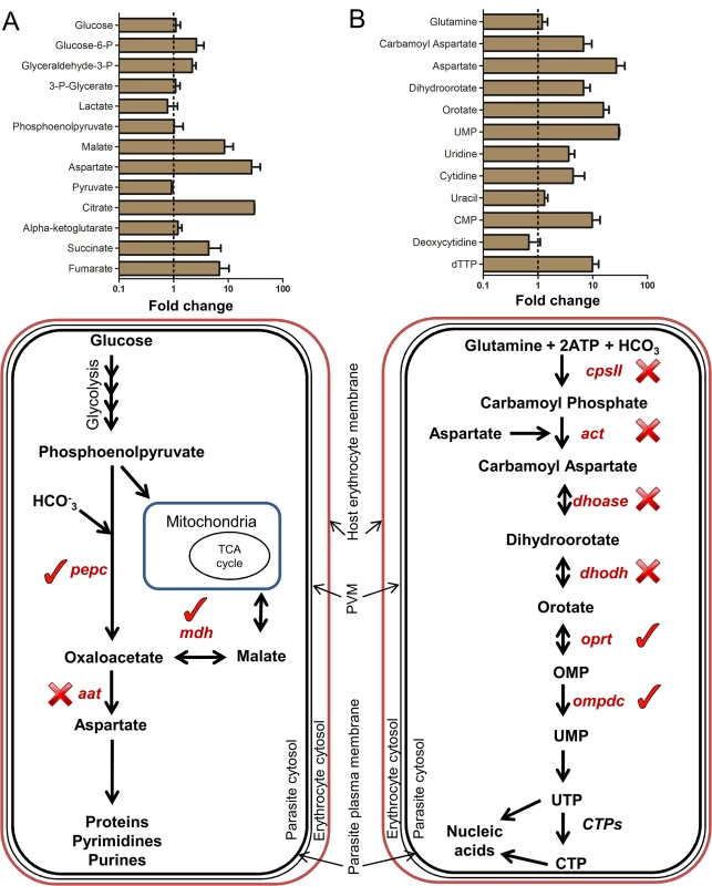
A. Top panel: Fold change of relative levels (peak intensities) of metabolites of carbon metabolism in rodent REP compared to wtEP. Dotted line indicates no change and error bars indicate R.S.D. (Relative Standard Deviation) of peak intensities from reticulocyte samples multiplied to the fold change values from n = 3 independent biological replicates. Bottom panel: Schematic representation of intermediary carbon metabolism (ICM) in Plasmodium cytosol. Genes marked with (✓) were deleted in P. berghei blood stages and the ones marked with (✕) could not be deleted even after repeated attempts. pepc: Phosphoenolpyruvate Carboxylase (PBANKA_101790), mdh: Malate Dehydrogenase (PBANKA_111770), aat: Aspartate Amino Transferase (PBANKA_030230). B. Top panel: Fold change of relative levels (peak intensities) of metabolites of pyrimidine biosynthesis in rodent REP compared to wtEP. Dotted line indicates no change and error bars indicate R.S.D. (Relative Standard Deviation) of peak intensities from reticulocyte samples multiplied to the fold change values from n = 3 independent biological replicates. Bottom panel: Schematic representation of pyrimidine biosynthesis pathway in Plasmodium cytosol. Genes marked with (✓) were deleted in P. berghei blood stages and the ones marked with (✕) could not be deleted even after repeated attempts. cpsII: Carbamoyl phosphate synthetase II (PBANKA_140670), act: Aspartate carbamoyltransferase (PBANKA_135770), dhoase: Dihydroorotase (PBANKA_133610), dhodh: Dihydroorotate dehydrogenase (PBANKA_010210), oprt: Orotate phosphoribosyltransferase (PBANKA_111240), ompdc: Orotidine 5′-monophosphate decarboxylase (PBANKA_050740). (Also see Fig B in S1 Text for gene deletion strategy and confirmation) Metabolites involved in TCA cycle and intermediary carbon metabolism (ICM), including malate and aspartate, were found to be substantially higher in REP compared to wtEP (Fig 2A). The elevated levels of these intermediates may possibly explain the previous observation that disruption of the TCA cycle in P. berghei blood stages through deletion of flavoprotein (Fp) subunit of the succinate dehydrogenase, pbsdha (PBANKA_051820), had little effect on parasite viability in blood stage forms, although ookinete development was impaired [49]. To further explore the possibility that P. berghei has potential access to the anapleurotic substrates of reticulocyte ICM, attempts were made to delete pepc, mdh and aat in P. berghei and assess the importance of these parasite enzymes throughout the life cycle (Fig 2A). P. berghei mutants lacking both pepc and mdh were generated (Fig B in S1 Text), while deletion of aat proved refractory. Both the pepc- and mdh- mutant parasites caused severe cerebral malaria in CD57/B6 mouse model with similar dynamics to wt parasites (Fig 3B). Interestingly, the growth of the pepc- mutant was compromised compared to wild type parasites, as the pepc- mutant, but not the mdh- mutant was overgrown by the wt parasite in an in vivo sensitive single host competitive growth assay (Fig 3A and C-A in S1 Text). The number of merozoites observed in mature schizont stages in both pepc- (17.02 ± 1.8) and mdh- (17.41 ± 1.7) mutants are similar to wt (17.4±1.8) (Fig C-C in S1 Text). Scrutiny of the growth phenotype detected in the pepc- mutants showed that they have a prolonged asexual cycle (4 h longer than wt) (p<0.05) (Fig C-B in S1 Text). The number of gametocytes formed in blood stages was also reduced in pepc- mutants by almost 50% but unaffected in mdh- (p>0.05) (Fig 3C) with no notable difference in male to female ratio in either mutant. Further phenotypic analyses showed reduction of exflagellation (pepc- mutants 84% less than wt, p<0.0005; mdh- mutants 56% less than wt, p<0.005) (Fig 3D). DNA replication in male gametocytes as observed by FACS analysis was reduced by 50% compared to wt at the 8 minute time point and further delayed taking up to 16 minutes to complete (Fig C-E and C-D in S1 Text). Ookinete development in in vitro cultures of pepc- mutants was also severely affected while in mdh- mutants, ookinetes were formed but the number was reduced by about 50% compared to wt (Fig 4A). To determine if this defect was sex specific, crosses of pepc- and mdh- were performed with P. berghei lines RMgm-348 (Pb270, p47-) which produces viable male gametes but non-viable female gametes and RMgm-15 (Pb137, p48/45-) which produces viable female gametes but non-viable male gametes [50]. Mutants of pepc- were found to produce severely reduced numbers of ookinetes in either cross suggesting that gametes of both genders are affected and that the activity of the protein is essential for viable gamete formation. This was not the case for mdh- mutants where although crossing experiments showed that lack of MDH protein affected both genders, they mimicked the parental phenotype producing 50% fewer mature ookinetes (Fig 4B). The pepc- parasites were defective in development within the mosquitoes, forming small numbers of oocysts in mosquito midguts and no salivary gland sporozoites. However, parasites lacking mdh could complete transmission through the mosquito and infect mice generating blood stage asexual forms in 48–72 hours similar to wt despite producing reduced numbers of oocysts when compared to wt (Fig 4C and 4D and D-A and D-B in S1 Text). Overall, these results suggest that two key enzymes in P. berghei ICM are at least partially redundant during stages of infection in which the parasites resides primarily in reticulocytes, but that they become essential as parasite differentiates and proliferates within other host or vector cell types.
Fig. 3. Phenotypic analyses of blood stage mutant P. berghei parasites. 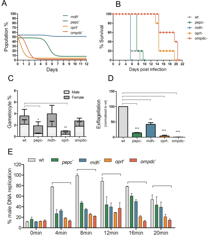
A. in vivo growth assay of mutants in mixed infections in competition with wild type parasites over 12 days. Coloured lines represent non-linear fit of percentage of mutant parasites in total parasite population. Data representative of n = 3 independent biological replicates. (Also see Fig C- A, B and C in S1 Text.) B. Lethality experiment in C57/B6 mice by wt and mutant P. berghei parasites. 104 parasites were injected intra-peritoneally in mice (n = 5) on day 0 and they were monitored for 21 days. The mice were culled humanely when they showed severe malaria pathology. All mutant parasites were found to be lethal to mice. C. Gametocyte conversions during blood stages in mutant P. berghei parasites over 5 days post infection. Data from 2 independent observed gametocyte conversion experiments are shown ± S.D. Gametocyte conversion was observed using a wt parent line which expresses GFP in male gametocytes and RFP in female gametocytes (RMgm-164). P. berghei mutants were generated in the same genetic background and analysed using FACS determining the number of gametocytes in infected blood. P-values: *p<0.05, **p<0.005, ***p<0.0005, paired two tailed t-test. D. Exflagellation (male gamete formation) in mutant P. berghei parasites normalised to wt in in vitro activation assay. The error is given as the SD of n = 3 independent biological replicates. P-values: **p<0.005, ***p<0.0005, paired two tailed t-test. E. Determination of DNA content of male gametocytes over 20 minutes post activation by FACS analysis in mutant P. berghei parasites normalised to wt. DNA content was determined in Hoechst-33258-stained MACS purified gametocytes. Before activation (0minutes) males show low DNA content with increasing amounts post activation reaching maximum levels between 8 to 12 minutes in wt. Data from 3 independent biological replicates are given ± S.D. P-values: **p<0.005, ***p<0.0005, unpaired two tailed t-test (also see Fig C- D in S1 Text). Fig. 4. Mosquito stage development of P. berghei mutant parasites (also see Fig D in S1 Text). 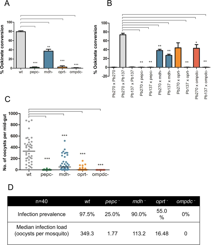
A. in vitro ookinete conversion of mutant P. berghei parasites as compared to wt. The error is given as the S.D. of n = 3 independent biological replicates. P-values: **p<0.005, ***p<0.0005, unpaired two tailed t-test. B. in vitro ookinete conversion assay to measure fertility of mutant P. berghei gametocytes. Fertility of mutant P. berghei gametocytes was analysed by their capacity to form ookinetes by crossing gametes with RMgm-348 (Pb270, p47-) which produces viable male gametes but non-viable female gametes and RMgm-15 (Pb137, p48/45-) which produces viable female gametes but non-viable male gametes. The error is given as the S.D. of n = 2 independent biological replicates. P-values: *p<0.05, **p<0.005, unpaired two tailed t-test. C. Number of mature oocysts at day 14 post infected blood feed in mosquito mid guts. n = 40 mosquitoes cumulative of two independent biological replicates. ***p<0.0005, unpaired two tailed t-test. D. Infection prevalence (percentage of observed mosquitoes found to be infected) and infection load (median of number of oocysts found per mosquito) in mutant P.berghei parasites compared to wt. Pyrimidine biosynthesis can be partially disrupted in reticulocyte-preferent P. berghei
Plasmodium spp. are heavily dependent on nucleic acid synthesis during blood stage asexual growth and either salvage (i.e. purines) or synthesize (i.e. pyrimidines) the requisite bases. A schematic representation of the pyrimidine biosynthesis pathway is given in Fig 2B. Five out of six enzymes of this pathway have been shown to be essential for P. falciparum growth in standard in vitro cultures, based on pharmacological studies [51]. Interestingly, most of these inhibitors are markedly less potent in the in vivo P. berghei model, a feature that has been attributed to reduced bio-availability of inhibitors in mice or apparent differences in target enzyme structures [52,53]. However, increased resistance to pyrimidine biosynthetic inhibitors could also reflect higher concentrations of pyrimidine precursors (bar glutamine) in the reticulocyte population selectively colonized by this species (Fig 2B) [16,51]. To investigate this possibility we attempted to delete in P. berghei 6 genes encoding enzymes involved in pyrimidine biosynthesis; carbamoyl phosphate synthetase II (cpsII) (PBANKA_140670), aspartate carbamoyltransferase (act) (PBANKA_135770), dihydroorotase (dhoase) (PBANKA_133610), dihydroorotate dehydrogenase (dhodh) (PBANKA_010210), orotate phosphoribosyltransferase (oprt) (PBANKA_111240) and orotidine 5′-monophosphate decarboxylase (ompdc) (PBANKA_050740). While the first four enzymes in this pathway were refractory to deletion, the last two enzymes in pyrimidine biosynthesis, orotate phosphoribosyltransferase (oprt) and orotidine 5′-monophosphate decarboxylase (ompdc) could be deleted (Fig B in S1 Text). The oprt- and ompdc- mutant parasites grew slowly (asexual cycle prolonged by approximately 4–5 hours compared to wt (p<0.05)), were rapidly outgrown in a competition growth assay with wt parasites (Fig 3A) and based on gray value-1 of staining intensity as observed by Giemsa staining (p<0.0005), seem to invade very young reticulocytes (Fig C-E in S1 Text). However, these infected reticulocytes could not be classified as CD71-high possibly due to the accelerated loss of the CD71 as observed with P. vivax infected reticulocytes [30]. Furthermore, both oprt- mutants (15.9 ± 2.0, p<0.0005) and ompdc- mutants (15.2 ± 2.5, p<0.0005) were found to generate, on average, significantly fewer merozoites than wt parasites (17.5 ± 1.8) per schizont (counted after completion of asexual cycle) (Fig C-C in S1 Text) and the asexual parasites also took longer to mature to schizonts (Fig C-B in S1 Text). Both mutants showed altered lethality in the C57/B6 mouse model as the mice infected with the mutants did not manifest the symptoms of experimental cerebral malaria (ECM) but died between days 14–20 as a result of severe anaemia and hyperparasitemia (Fig 3B). The process of transmission was also affected by the loss of ompdc and oprt. Gametocytemia was significantly reduced only in oprt- parasites (Fig 3C) but no change was seen in male - female ratio. Exflagellation (the production of mature male gametes) was found to be severely affected in oprt- and completely blocked in ompdc- parasites (Fig 3D) and DNA replication during male gametogenesis was severely reduced (Fig 3E). Consistent with the defects in male gametogenesis, very few ookinetes were formed in in vitro cultures in oprt- parasites and no ookinetes were observed in ompdc- (Fig 4A). Genetic crosses of oprt- and ompdc- mutants were performed as above with P. berghei lines RMgm-348 and RMgm-15 which showed that viable male gametes (from RMgm-348) were able to rescue the ookinete conversion defect in both mutant lines suggesting that formation of male gametes is impaired in both oprt- and ompdc- mutant parasites while female gametes remain unaffected (Fig 4B). Infectivity to the mosquito was significantly reduced in oprt- and completely blocked in ompdc- mutants as seen by observing oocysts in infected mosquito midguts and salivary gland sporozoites (Fig 4C and 4D and D-C and D-D in S1 Text) and infection to naïve mice was found to be completely blocked. However, when ookinetes from p47- x oprt- or ompdc- crosses were fed to mosquitoes, they failed to develop into mature oocysts (Fig E in S1 Text) hence, did not complete sporogony indicating that lack of both oprt and ompdc in the female lineage results in an allelic insufficiency in a growing oocyst.
We also tested the effect of a previously published inhibitor of pyrimidine biosynthesis 5-fluoroorotate (5FOA) [54] on asexual growth of both P. falciparum and P. berghei. The comparisons were carried out in vitro to prevent bioavailability of the inhibitor confounding in vivo data in mice. We tested the activity and found that the IC50 value of 5FOA in vitro was almost 90-fold higher in P. berghei (32.2 ± 0.9 nM) compared to P. falciparum (0.37 ± 0.01 nM) (Fig 5). A dihydroartemisinin control showed no major difference in inhibition between P. berghei (6.6 ± 0.1 nM) and P. falciparum (2.8 ± 0.2 nM). These data strongly suggest that P. berghei can access pyrimidine precursors from the reticulocyte and are consistent with a role of host cell metabolism in the differential activity of 5FOA, although differences in sensitivity of P. falciparum [55] and P. berghei [56] thymidylate synthase or differences in drug uptake could also contribute to the differential lethality.
Fig. 5. P. berghei and P. falciparum inhibition by dihydroartemisinin (DHA) and 5-fluoroorotic acid (5FOA) in vitro. 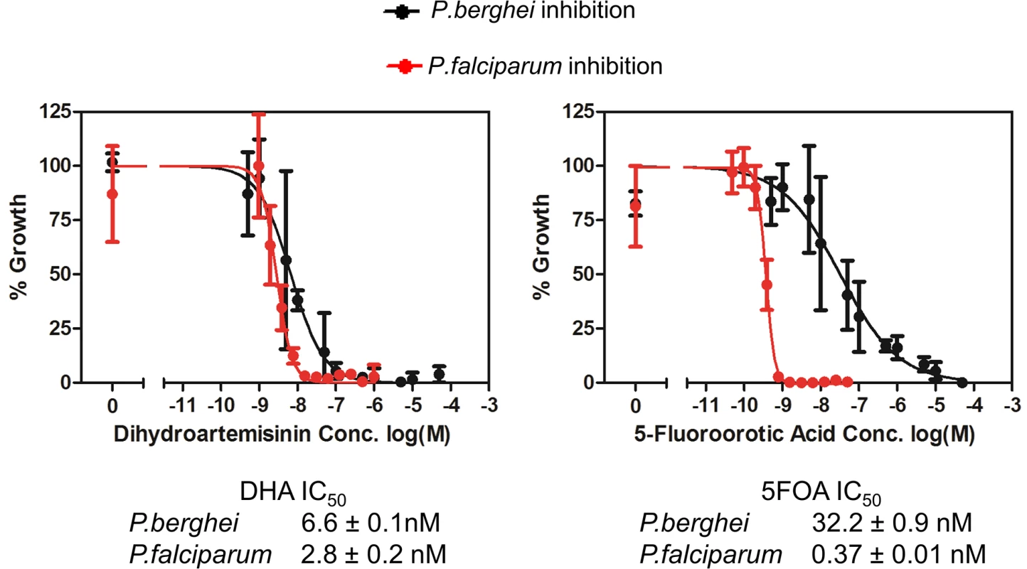
Error bars indicate S.D. from n = 3 biological replicates. Discussion
These metabolomics analyses clearly showed that reticulocytes have a much more complex metabolome than mature erythrocytes, adding to previously well documented changes that occur in both organelle complement and protein expression levels during reticulocyte maturation in peripheral circulation [34,35]. Key metabolic processes that were found to be elevated in REP but absent or highly reduced in wtEP included the TCA cycle and associated intermediary carbon metabolism, nucleic acid metabolism, phospholipid metabolism, fatty acid catabolism and glutathione metabolism. The down-regulation of these host pathways in wtEP may explain why several of the corresponding pathways in asexual blood stages of P. falciparum appear to be essential in vitro. Conversely, we predicted that malaria parasites infectious to both humans (P. vivax) and rodents (P. berghei) which exhibit a strong tropism for reticulocytes rather than mature erythrocytes may be more tolerant to loss of key metabolic pathways because of redundancy with host pathways. Our data strongly suggest that reticulocytes do indeed provide a highly enriched host cell niche for Plasmodium, with important implications for drug discovery strategies. Although the reticulocyte offers a very different nutrient resource compared to the mature erythrocyte, there has been apparently little change in the metabolic capacity of P. vivax and P. berghei. Our in silico comparison (S3 Table) reveals that P. vivax and P. berghei have 98% and 97% orthology, respectively, to P. falciparum genes annotated with metabolic pathway information (434 genes) on PlasmoDB.
The enriched reticulocyte metabolome supports growth of metabolically compromised P. berghei
Although a small number of metabolomics studies have been undertaken on erythrocytes, including a comparison of normal and HbSS erythrocytes, the number and range of metabolites detected in these studies were relatively small (20–90) [39,40]. Here we used complementary LC-MS and GS-MS analytical platforms to maximise coverage, generating the most comprehensive coverage of REP and wtEP undertaken to date (333 metabolites). These studies revealed a much higher degree of metabolic complexity in reticulocytes compared to mature erythrocytes covering nearly all major pathways in central carbon metabolism. Whilst glycolysis is the main pathway for carbon metabolism in erythrocytes [36], both human [57] and rodent [58] erythrocytes retain a residual proteomic signature of TCA cycle and ICM enzymes and our metabolomics data suggests that these pathways are much more active in reticulocytes, leading to elevated levels of TCA intermediates (including citrate, malate) and ICM products (e.g. aspartate). The functional significance of increased metabolic complexity in reticulocytes was subsequently tested by generating P. berghei mutants with specific defects in metabolism showing that the increased availability of complementary metabolites in reticulocytes can explain the non-essential nature of the P. berghei pepc and mdh genes, which are involved in regulating intracellular levels of oxaloacetate and malate. In contrast, PEPC is essential for normal intra-erythrocytic survival of P. falciparum in vitro, although this can be bypassed by malate supplementation of P. falciparum infected mature erythrocytes [42]. It should be noted that whilst the P. berghei pepc- mutant retained its virulence, it still showed a significant growth defect compared with wild type parasites (similar to the P. falciparum mutant [42]) resulting at least in part from a prolongation of the asexual blood stage cycle as revealed by our sensitive single host competitive growth assay. It would be interesting to use this assay to compare asexual growth dynamics of other available metabolic mutants such as the pbsdha- with wild type which might reveal additional defects to those reported [49]. The P. berghei pepc- mutant also failed to complete transmission through mosquitoes as a result of defects in gametocyte production, male gamete formation, female gamete viability resulting in trace oocyst formation and failure to enter sporogony, which extends our understanding of the importance of this metabolic enzyme for parasite development beyond the asexual blood stages previously investigated [42]. A possible explanation for this phenotype is that the pepc- mutant is unable to by-pass the need for de novo synthesized aspartate for nucleotide biosynthesis by salvage from different host cells during its sexual and asexual life cycle (Fig 2A). The demonstration of pbpepc- growth in reticulocytes suggests that the equivalent P. falciparum mutant might be a suitable candidate for an attenuated slow growing parasite vaccine that would permit generation of significant anti-parasitic immune responses.
In line with this suggestion, we were unable to delete pbaat which is also required for de novo synthesis of aspartate. The essential nature of pbaat suggests that either the apparently higher levels of aspartate in reticulocytes are insufficient to meet the demands of a growing asexual stage parasite or that, as in P. falciparum intra-erythrocytic stages, P. berghei is not readily able to access host cytoplasmic pools of aspartate [59]. Production of aspartate in Plasmodium pepc- mutants can still be achieved through generation of the oxaloacetic acid precursor by mitochondrial malate: quinone oxidoreductase (MQO) or the reverse reaction of cytosolic MDH. However, this is apparently a suboptimal solution for the pepc- parasite resulting in slow growth in the blood stage and failure to develop in the mosquito. Plasmodium AAT can also generate methionine from aspartate, glutamate and other amino acids which can act as effective amino group donors [60] and regulate glutamine/glutamate metabolism. These functions may not be rescued by simple aspartate salvage from the host and further support the essentiality of aat as a key enzyme for the parasite and a possible drug target.
The P. falciparum gene encoding MDH has proved refractory to deletion under any circumstances so far, suggesting that it is essential for these parasites. In marked contrast, the P. berghei mutants lacking pbmdh were readily generated, suggesting that this species may scavenge reticulocyte pools of malate or other intermediates in the TCA cycle. The pbmdh mutant exhibited a very modest growth phenotype and was able to develop into mosquito infective stages, although it produced 30% fewer oocysts than wt parasites. The continued viability of the pbmdh- mutants during transmission in the absence of reticulocyte-based compensatory sources of the metabolite can be explained by continued TCA derived production of malate and NADH+ H+ reducing equivalents given the increased flux through the TCA metabolism in gametocytes and probably later sexual stages [7,12]. Conditional silencing or disruption of pfmdh or degradation of PfMDH in mature gametocytes or later stages of P. falciparum would establish if MDH is required for transmission of the human parasite and that the essential nature of this enzyme is merely blood stage specific.
P. berghei is partially capable of pyrimidine salvage in highly immature reticulocytes
Plasmodium spp. salvage their purine requirements from the host cell, but retain the ability to synthesise pyrimidines [61]. Purine nucleosides are taken up by the parasite PfNT1 and other, as yet, unidentified AMP transporters [62] after they are delivered to the parasitophorous vacuole via the action of erythrocyte nucleoside transporters [51,63] and a non-selective transport process [61,64]. In contrast, while other Apicomplexans (i.e. Cryptosporidium spp., Toxoplasma spp.) retain the capacity to salvage pyrimidines [16], Plasmodium spp. are thought to lack enzymes required for host pyrimidine salvage [44]. Although, Plasmodium proteins have been implicated in transporting some pyrimidine precursors [65,66], presumably due to very limited availability of pyrimidines in the host cell, Plasmodium parasites have been thought to be completely dependent on de novo pyrimidine synthesis for growth in asexual stages [51].
The survival of both oprt-and ompdc- mutants could be the result of two possibilities that are not mutually exclusive. The first possibility is that the mutants directly utilize reticulocyte pools of pyrimidines which are nonetheless limiting leading to a reduction in number of merozoites produced. Alternatively, mutant parasites could synthesize orotate which is secreted into the host cytoplasm and converted to UMP by host UMP synthase before being salvaged. Both outcomes require transport of nucleosides or nucleotides from the host cytoplasm to the parasite and how this is achieved is not clear. Both pyrimidine biosynthesis mutants survive only in the youngest reticulocytes which might reflect either adequacy of supply of host UMP (or derivatives) or the capacity of the youngest reticulocytes to convert parasite-derived orotate. Indeed enzymes involved in the later stages of pyrimidine biosynthesis, nucleoside diphosphate kinase B, CTP synthase and ribonucleotide reductase large subunit have been identified in rodent and human erythrocytes [57,58]. The possibility that host pyrimidine enzymes may have redundant functions with the parasite enzymes catalysing late steps in pyrimidine biosynthesis is supported by the apparent essentiality of the P. berghei genes encoding the first four steps of pyrimidine biosynthesis. A simplified illustration of life cycle stages of P. berghei development showing the characteristics of mutant parasites at various points in the life cycle is shown in Fig G in S1 Text.
Glutathione biosynthesis is elevated in reticulocytes
The REP metabolome also explains other species-specific differences between P. berghei and P. falciparum. Glutathione biosynthesis occurs in erythrocytes [67] and the enzymes for this pathway have been shown to be present in both human [57] and rodent [58] erythrocytes. Plasmodium employs its own glutathione redox system [68] to counter oxidative stress (Fig F-A in S1 Text). Both ɣ-glutamylcysteine synthetase (ɣ-gcs) and glutathione synthetase (gs) are essential for parasite survival in P. falciparum [69] yet ɣ-gcs and glutathione reductase (gr) can be deleted in P. berghei and intra-erythrocytic asexual growth is unaffected although mosquito stage development is arrested at the oocyst stage [70,71]. The REP and wtEP metabolomes demonstrated that the levels of glutathione synthesis intermediates were higher in reticulocytes than in mature erythrocytes (Fig F-B in S1 Text) providing a mechanistic explanation for the normal growth of P. berghei ɣ-gcs and gr mutant asexual stages in reticulocytes. Also, the inhibitor of ɣ-gcs, buthionine sulphoximine (BSO) inhibits P. falciparum growth with an IC50 value of ~60 μM [69] yet concentrations as high as 500 μM BSO in vitro had no inhibitory effect on P. berghei parasites in in vitro cultures (Fig F-C in S1 Text) consistent with the reticulocyte mediated rescue of chemical disruption of the glutathione synthesis pathway in P. berghei, although it has not been investigated whether there is a difference in BSO sensitivity against the mouse or P. berghei ɣ-gcs compared to P.falciparum or human enzymes.
Host cell metabolism can ameliorate the impact of different drug treatments
Enzymes involved in Plasmodium intermediary carbon metabolism [12,42] and pyrimidine biosynthesis [51] are considered attractive targets for drug development. The metabolome surveys and drug inhibition data presented here suggest that caution should be used before extrapolating conclusions regarding gene essentiality in reticulocyte preferent parasites such as P. berghei as part of any drug discovery pathway that has been based initially upon screens in mature erythrocytes. Bioavailability in mouse models and/or drug penetration into the reticulocyte and difference in target enzyme structures between species have been proposed as reasons for the relative ineffectiveness of drugs when tested in vivo using P. berghei [52,53]. An alternative view is that the reticulocyte metabolome (at least in part) provides a reservoir of metabolites downstream of the point of action of a drug rendering the drug less effective. This has a number of consequences:
Good drug candidates for P. falciparum might be (already have been) eliminated from further development due to adverse and misleading data from in vivo P. berghei testing.
Alternative mature erythrocyte preferent rodent parasites (e.g. P. yoelii YM) [72] might provide a more accurate in vivo model in which to test drug candidates for P. falciparum malaria.
P. berghei could provide an accurate in vivo model for the development of drugs against reticulocyte preferent parasites such as P. vivax, and this warrants further investigation.
Recrudescence of P. falciparum infection during treatment with anti-malarials that target parasite metabolism (anti-metabolites) might result from their survival in a ‘protective’ reticulocyte niche, the existence of which cannot be predicted by standard in vitro cultures.
Reticulocyte resident parasites exposed to anti-metabolites at levels determined by in vitro culture in mature erythrocytes would be sub-optimal with the subsequent risk of being more conducive for the selection of drug resistant progeny.
Materials and Methods
Metabolomics of rodent erythrocytes
Rodent reticulocyte enrichment was done in rats by administering phenylhydrazine-HCl dissolved in 0.9% NaCl (w/v) at a dose of 100 mg/kg body weight and collecting reticulocyte enriched peripheral blood on day 5 post injection. Metabolite extraction was done as using chloroform/methanol/water (1 : 3:1 v/v) and samples were analysed using LC-MS and GC-MS. See S1 supplementary materials and methods for details.
Metabolomics of human CD34+ stem cell grown erythrocytes
CD34+ cells obtained from blood from human volunteers were cultured in a three-stage protocol based on the methods of [41]. Cultured reticulocytes and mature erythrocytes from matching donors were used for metabolite extraction with chloroform/methanol/water (1 : 3:1 v/v) and samples were analyzed using LC-MS. See S1 supplementary materials and methods for details.
P. berghei methods
Infection of laboratory mice, asexual culture of P. berghei stages and generation of knockout parasites was done as before [73]. Asexual growth competition assay was done by mixing wt and mutant parasites expressing different fluorescent markers and injecting them intravenously into recipient mice and monitoring the growth of the two populations by flow cytometry as done before [74]. Lethality of mutant P. berghei parasites was checked by injecting infected RBCs (104) into C57/B6 mice and monitoring parasitaemia, disease pathology and mortality over 21 days. Gametocyte conversion was monitored by flow cytometry in mutants generated in parent line (820cl1m1cl1) expressing GFP in male gametocytes and RFP in female gametocytes [75]. DNA quantification during exflagellation was also monitored by flow cytometry in mutant P. berghei parasites. Development of ookinetes in wild type, mutants and sexual crosses was observed in standard in vitro cultures maintained at 21°C. Mosquito transmission experiments were done in 5–8 days old mosquitoes used for infected blood feeds at 21°C and monitored for oocyst and sporozoite development. See S1 supplementary materials and methods for details.
Determination of IC50 value of inhibitors in vitro
Inhibitors were used to perform in vitro drug susceptibility tests in standard cultures of synchronized P. berghei and P. falciparum blood stages. For testing P. berghei inhibiton, inhibitors were used at increasing concentrations to culture ring stage P. berghei for 24 hours and parasite development to schizont stage was analyzed by flow cytometry after staining iRBCs with DNA-specific dye Hoechst-33258. P. falciparum 3D7 strain was used for determining IC50 values of inhibitors in in vitro cultures by measuring 3H-Hypoxanthine incorporation in the presence of inhibitors in increasing concentrations. See S1 supplementary materials and methods for details.
Ethics statement
All animal work was approved by the University of Glasgow’s Animal Welfare and Ethical Review Body and by the UK’s Home Office (PPL 60/4443). The animal care and use protocol complied with the UK Animals (Scientific Procedures) Act 1986 as amended in 2012 and with European Directive 2010/63/EU on the Protection of Animals Used for Scientific Purposes. Blood from human volunteers was supplied by the Australian Red Cross Blood Service and experiments were approved by the Walter and Eliza Hall Institute Human Research Ethics Committee, Australia. As a part of standard Australian Red Cross Blood Service practice, blood was collected from healthy donors who were informed about this study and potential risks to them and gave written consent when they donated blood.
Supporting Information
Zdroje
1. Bozdech Z, Llinas M, Pulliam BL, Wong ED, Zhu J, et al. (2003) The transcriptome of the intraerythrocytic developmental cycle of Plasmodium falciparum. PLoS Biol 1: E5. 12929205
2. Llinas M, del Portillo HA (2005) Mining the malaria transcriptome. Trends Parasitol 21 : 350–352. 15979411
3. Khan SM, Franke-Fayard B, Mair GR, Lasonder E, Janse CJ, et al. (2005) Proteome analysis of separated male and female gametocytes reveals novel sex-specific Plasmodium biology. Cell 121 : 675–687. 15935755
4. Hall N, Karras M, Raine JD, Carlton JM, Kooij TW, et al. (2005) A comprehensive survey of the Plasmodium life cycle by genomic, transcriptomic, and proteomic analyses. Science 307 : 82–86. 15637271
5. Olszewski KL, Morrisey JM, Wilinski D, Burns JM, Vaidya AB, et al. (2009) Host-parasite interactions revealed by Plasmodium falciparum metabolomics. Cell Host Microbe 5 : 191–199. doi: 10.1016/j.chom.2009.01.004 19218089
6. Kafsack BF, Llinas M (2010) Eating at the table of another: metabolomics of host-parasite interactions. Cell Host Microbe 7 : 90–99. doi: 10.1016/j.chom.2010.01.008 20159614
7. Macrae JI, Dixon MW, Dearnley MK, Chua HH, Chambers JM, et al. (2013) Mitochondrial metabolism of sexual and asexual blood stages of the malaria parasite Plasmodium falciparum. BMC Biol 11 : 67. doi: 10.1186/1741-7007-11-67 23763941
8. Sana TR, Gordon DB, Fischer SM, Tichy SE, Kitagawa N, et al. (2013) Global mass spectrometry based metabolomics profiling of erythrocytes infected with Plasmodium falciparum. PLoS One 8: e60840. doi: 10.1371/journal.pone.0060840 23593322
9. Booden T, Hull RW (1973) Nucleic acid precursor synthesis by Plasmodium lophurae parasitizing chicken erythrocytes. Exp Parasitol 34 : 220–228. 4744840
10. Sherman IW (1977) Amino acid metabolism and protein synthesis in malarial parasites. Bull World Health Organ 55 : 265–276. 338183
11. Homewood CA (1977) Carbohydrate metabolism of malarial parasites. Bull World Health Organ 55 : 229–235. 338181
12. Oppenheim RD, Creek DJ, Macrae JI, Modrzynska KK, Pino P, et al. (2014) BCKDH: The Missing Link in Apicomplexan Mitochondrial Metabolism Is Required for Full Virulence of Toxoplasma gondii and Plasmodium berghei. PLoS Pathog 10: e1004263. doi: 10.1371/journal.ppat.1004263 25032958
13. Holz GG Jr. (1977) Lipids and the malarial parasite. Bull World Health Organ 55 : 237–248. 412602
14. Dechamps S, Shastri S, Wengelnik K, Vial HJ (2010) Glycerophospholipid acquisition in Plasmodium—a puzzling assembly of biosynthetic pathways. Int J Parasitol 40 : 1347–1365. doi: 10.1016/j.ijpara.2010.05.008 20600072
15. Barrett MP (1997) The pentose phosphate pathway and parasitic protozoa. Parasitol Today 13 : 11–16. 15275160
16. Hyde JE (2007) Targeting purine and pyrimidine metabolism in human apicomplexan parasites. Curr Drug Targets 8 : 31–47. 17266529
17. Macedo CS, Schwarz RT, Todeschini AR, Previato JO, Mendonca-Previato L (2010) Overlooked post-translational modifications of proteins in Plasmodium falciparum: N - and O-glycosylation—a review. Mem Inst Oswaldo Cruz 105 : 949–956. 21225189
18. Landfear SM (2011) Nutrient transport and pathogenesis in selected parasitic protozoa. Eukaryot Cell 10 : 483–493. doi: 10.1128/EC.00287-10 21216940
19. Martin RE, Ginsburg H, Kirk K (2009) Membrane transport proteins of the malaria parasite. Mol Microbiol 74 : 519–528. doi: 10.1111/j.1365-2958.2009.06863.x 19796339
20. Maier AG, Duraisingh MT, Reeder JC, Patel SS, Kazura JW, et al. (2003) Plasmodium falciparum erythrocyte invasion through glycophorin C and selection for Gerbich negativity in human populations. Nat Med 9 : 87–92. 12469115
21. Duraisingh MT, Maier AG, Triglia T, Cowman AF (2003) Erythrocyte-binding antigen 175 mediates invasion in Plasmodium falciparum utilizing sialic acid-dependent and-independent pathways. Proc Natl Acad Sci U S A 100 : 4796–4801. 12672957
22. Mayer DC, Cofie J, Jiang L, Hartl DL, Tracy E, et al. (2009) Glycophorin B is the erythrocyte receptor of Plasmodium falciparum erythrocyte-binding ligand, EBL-1. Proc Natl Acad Sci U S A 106 : 5348–5352. doi: 10.1073/pnas.0900878106 19279206
23. Crosnier C, Bustamante LY, Bartholdson SJ, Bei AK, Theron M, et al. (2011) Basigin is a receptor essential for erythrocyte invasion by Plasmodium falciparum. Nature 480 : 534–537. doi: 10.1038/nature10606 22080952
24. Tham WH, Healer J, Cowman AF (2012) Erythrocyte and reticulocyte binding-like proteins of Plasmodium falciparum. Trends Parasitol 28 : 23–30. doi: 10.1016/j.pt.2011.10.002 22178537
25. Harvey KL, Gilson PR, Crabb BS (2012) A model for the progression of receptor-ligand interactions during erythrocyte invasion by Plasmodium falciparum. Int J Parasitol 42 : 567–573. doi: 10.1016/j.ijpara.2012.02.011 22710063
26. Wright GJ, Rayner JC (2014) Plasmodium falciparum erythrocyte invasion: combining function with immune evasion. PLoS Pathog 10: e1003943. doi: 10.1371/journal.ppat.1003943 24651270
27. Pasvol G, Weatherall DJ, Wilson RJ (1980) The increased susceptibility of young red cells to invasion by the malarial parasite Plasmodium falciparum. Br J Haematol 45 : 285–295. 7002199
28. Galinski MR, Medina CC, Ingravallo P, Barnwell JW (1992) A reticulocyte-binding protein complex of Plasmodium vivax merozoites. Cell 69 : 1213–1226. 1617731
29. Barnwell JW, Nichols ME, Rubinstein P (1989) In vitro evaluation of the role of the Duffy blood group in erythrocyte invasion by Plasmodium vivax. J Exp Med 169 : 1795–1802. 2469769
30. Malleret B, Li A, Zhang R, Tan KS, Suwanarusk R, et al. (2014) Plasmodium vivax: restricted tropism and rapid remodelling of CD71 positive reticulocytes. Blood.
31. Cromer D, Evans KJ, Schofield L, Davenport MP (2006) Preferential invasion of reticulocytes during late-stage Plasmodium berghei infection accounts for reduced circulating reticulocyte levels. Int J Parasitol 36 : 1389–1397. 16979643
32. Janse CJ, Waters AP (1995) Plasmodium berghei: the application of cultivation and purification techniques to molecular studies of malaria parasites. Parasitol Today 11 : 138–143. 15275357
33. Chen K, Liu J, Heck S, Chasis JA, An X, et al. (2009) Resolving the distinct stages in erythroid differentiation based on dynamic changes in membrane protein expression during erythropoiesis. Proc Natl Acad Sci U S A 106 : 17413–17418. doi: 10.1073/pnas.0909296106 19805084
34. Gronowicz G, Swift H, Steck TL (1984) Maturation of the reticulocyte in vitro. J Cell Sci 71 : 177–197. 6097593
35. Liu J, Guo X, Mohandas N, Chasis JA, An X (2010) Membrane remodeling during reticulocyte maturation. Blood 115 : 2021–2027. doi: 10.1182/blood-2009-08-241182 20038785
36. Chapman RG, Hennessey MA, Waltersdorph AM, Huennekens FM, Gabrio BW (1962) Erythrocyte metabolism. V. Levels of glycolytic enzymes and regulation of glycolysis. J Clin Invest 41 : 1249–1256. 13878200
37. Stromme JH, Eldjarn L (1962) The role of the pentose phosphate pathway in the reduction of methaemoglobin in human erythrocytes. Biochem J 84 : 406–410. 13917855
38. Dajani RM, Orten JM (1958) [A study of the citric acid cycle in erythrocytes]. J Biol Chem 231 : 913–924. 13539026
39. Malleret B, Xu F, Mohandas N, Suwanarusk R, Chu C, et al. (2013) Significant biochemical, biophysical and metabolic diversity in circulating human cord blood reticulocytes. PLoS One 8: e76062. doi: 10.1371/journal.pone.0076062 24116088
40. Darghouth D, Koehl B, Madalinski G, Heilier JF, Bovee P, et al. (2011) Pathophysiology of sickle cell disease is mirrored by the red blood cell metabolome. Blood 117: e57–66. doi: 10.1182/blood-2010-07-299636 21135259
41. Douay L, Giarratana MC (2009) Ex vivo generation of human red blood cells: a new advance in stem cell engineering. Methods Mol Biol 482 : 127–140. doi: 10.1007/978-1-59745-060-7_8 19089353
42. Storm J, Sethia S, Blackburn GJ, Chokkathukalam A, Watson DG, et al. (2014) Phosphoenolpyruvate carboxylase identified as a key enzyme in erythrocytic Plasmodium falciparum carbon metabolism. PLoS Pathog 10: e1003876. doi: 10.1371/journal.ppat.1003876 24453970
43. Ginsburg H (2006) Progress in in silico functional genomics: the malaria Metabolic Pathways database. Trends Parasitol 22 : 238–240. 16707276
44. Gardner MJ, Hall N, Fung E, White O, Berriman M, et al. (2002) Genome sequence of the human malaria parasite Plasmodium falciparum. Nature 419 : 498–511. 12368864
45. Valentine WN, Paglia DE (1980) Erythrocyte disorders of purine and pyrimidine metabolism. Hemoglobin 4 : 669–681. 6254919
46. Rebouche CJ, Paulson DJ (1986) Carnitine metabolism and function in humans. Annu Rev Nutr 6 : 41–66. 3524622
47. Bulusu V, Jayaraman V, Balaram H (2011) Metabolic fate of fumarate, a side product of the purine salvage pathway in the intraerythrocytic stages of Plasmodium falciparum. J Biol Chem 286 : 9236–9245. doi: 10.1074/jbc.M110.173328 21209090
48. Wrenger C, Muller IB, Schifferdecker AJ, Jain R, Jordanova R, et al. (2011) Specific inhibition of the aspartate aminotransferase of Plasmodium falciparum. J Mol Biol 405 : 956–971. doi: 10.1016/j.jmb.2010.11.018 21087616
49. Hino A, Hirai M, Tanaka TQ, Watanabe Y, Matsuoka H, et al. (2012) Critical roles of the mitochondrial complex II in oocyst formation of rodent malaria parasite Plasmodium berghei. Journal of biochemistry 152 : 259–268. doi: 10.1093/jb/mvs058 22628552
50. van Dijk MR, Janse CJ, Thompson J, Waters AP, Braks JA, et al. (2001) A central role for P48/45 in malaria parasite male gamete fertility. Cell 104 : 153–164. 11163248
51. Cassera MB, Zhang Y, Hazleton KZ, Schramm VL (2011) Purine and pyrimidine pathways as targets in Plasmodium falciparum. Curr Top Med Chem 11 : 2103–2115. 21619511
52. Gujjar R, Marwaha A, El Mazouni F, White J, White KL, et al. (2009) Identification of a metabolically stable triazolopyrimidine-based dihydroorotate dehydrogenase inhibitor with antimalarial activity in mice. J Med Chem 52 : 1864–1872. doi: 10.1021/jm801343r 19296651
53. Phillips MA, Gujjar R, Malmquist NA, White J, El Mazouni F, et al. (2008) Triazolopyrimidine-based dihydroorotate dehydrogenase inhibitors with potent and selective activity against the malaria parasite Plasmodium falciparum. J Med Chem 51 : 3649–3653. doi: 10.1021/jm8001026 18522386
54. Rathod PK, Khatri A, Hubbert T, Milhous WK (1989) Selective activity of 5-fluoroorotic acid against Plasmodium falciparum in vitro. Antimicrob Agents Chemother 33 : 1090–1094. 2675756
55. Hekmat-Nejad M, Rathod PK (1996) Kinetics of Plasmodium falciparum thymidylate synthase: interactions with high-affinity metabolites of 5-fluoroorotate and D1694. Antimicrob Agents Chemother 40 : 1628–1632. 8807052
56. Pattanakitsakul SN, Ruenwongsa P (1984) Characterization of thymidylate synthetase and dihydrofolate reductase from Plasmodium berghei. Int J Parasitol 14 : 513–520. 6392123
57. Pasini EM, Kirkegaard M, Mortensen P, Lutz HU, Thomas AW, et al. (2006) In-depth analysis of the membrane and cytosolic proteome of red blood cells. Blood 108 : 791–801. 16861337
58. Pasini EM, Kirkegaard M, Salerno D, Mortensen P, Mann M, et al. (2008) Deep coverage mouse red blood cell proteome: a first comparison with the human red blood cell. Mol Cell Proteomics 7 : 1317–1330. doi: 10.1074/mcp.M700458-MCP200 18344233
59. Martin RE, Kirk K (2007) Transport of the essential nutrient isoleucine in human erythrocytes infected with the malaria parasite Plasmodium falciparum. Blood 109 : 2217–2224. 17047158
60. Berger LC, Wilson J, Wood P, Berger BJ (2001) Methionine regeneration and aspartate aminotransferase in parasitic protozoa. J Bacteriol 183 : 4421–4434. 11443076
61. Downie MJ, Kirk K, Mamoun CB (2008) Purine salvage pathways in the intraerythrocytic malaria parasite Plasmodium falciparum. Eukaryot Cell 7 : 1231–1237. doi: 10.1128/EC.00159-08 18567789
62. Cassera MB, Hazleton KZ, Riegelhaupt PM, Merino EF, Luo M, et al. (2008) Erythrocytic adenosine monophosphate as an alternative purine source in Plasmodium falciparum. J Biol Chem 283 : 32889–32899. doi: 10.1074/jbc.M804497200 18799466
63. Quashie NB, Ranford-Cartwright LC, de Koning HP (2010) Uptake of purines in Plasmodium falciparum-infected human erythrocytes is mostly mediated by the human equilibrative nucleoside transporter and the human facilitative nucleobase transporter. Malar J 9 : 36. doi: 10.1186/1475-2875-9-36 20113503
64. Desai SA, Krogstad DJ, McCleskey EW (1993) A nutrient-permeable channel on the intraerythrocytic malaria parasite. Nature 362 : 643–646. 7681937
65. Parker MD, Hyde RJ, Yao SY, McRobert L, Cass CE, et al. (2000) Identification of a nucleoside/nucleobase transporter from Plasmodium falciparum, a novel target for anti-malarial chemotherapy. Biochem J 349 : 67–75. 10861212
66. Frame IJ, Merino EF, Schramm VL, Cassera MB, Akabas MH (2012) Malaria parasite type 4 equilibrative nucleoside transporters (ENT4) are purine transporters with distinct substrate specificity. Biochem J 446 : 179–190. doi: 10.1042/BJ20112220 22670848
67. Majerus PW, Brauner MJ, Smith MB, Minnich V (1971) Glutathione synthesis in human erythrocytes. II. Purification and properties of the enzymes of glutathione biosynthesis. The Journal of clinical investigation 50 : 1637–1643. 5097571
68. Muller S (2004) Redox and antioxidant systems of the malaria parasite Plasmodium falciparum. Molecular microbiology 53 : 1291–1305. 15387810
69. Patzewitz EM, Wong EH, Muller S (2012) Dissecting the role of glutathione biosynthesis in Plasmodium falciparum. Molecular microbiology 83 : 304–318. doi: 10.1111/j.1365-2958.2011.07933.x 22151036
70. Pastrana-Mena R, Dinglasan RR, Franke-Fayard B, Vega-Rodriguez J, Fuentes-Caraballo M, et al. (2010) Glutathione reductase-null malaria parasites have normal blood stage growth but arrest during development in the mosquito. The Journal of biological chemistry 285 : 27045–27056. doi: 10.1074/jbc.M110.122275 20573956
71. Vega-Rodriguez J, Franke-Fayard B, Dinglasan RR, Janse CJ, Pastrana-Mena R, et al. (2009) The glutathione biosynthetic pathway of Plasmodium is essential for mosquito transmission. PLoS pathogens 5: e1000302. doi: 10.1371/journal.ppat.1000302 19229315
72. Walliker D, Sanderson A, Yoeli M, Hargreaves BJ (1976) A genetic investigation of virulence in a rodent malaria parasite. Parasitology 72 : 183–194. 1264490
73. Janse CJ, Ramesar J, Waters AP (2006) High-efficiency transfection and drug selection of genetically transformed blood stages of the rodent malaria parasite Plasmodium berghei. Nat Protoc 1 : 346–356. 17406255
74. Sinha A, Hughes KR, Modrzynska KK, Otto TD, Pfander C, et al. (2014) A cascade of DNA-binding proteins for sexual commitment and development in Plasmodium. Nature 507 : 253–257. doi: 10.1038/nature12970 24572359
75. Ponzi M, Siden-Kiamos I, Bertuccini L, Curra C, Kroeze H, et al. (2009) Egress of Plasmodium berghei gametes from their host erythrocyte is mediated by the MDV-1/PEG3 protein. Cell Microbiol 11 : 1272–1288. doi: 10.1111/j.1462-5822.2009.01331.x 19438517
Štítky
Hygiena a epidemiologie Infekční lékařství Laboratoř
Článek Clearance of Pneumococcal Colonization in Infants Is Delayed through Altered Macrophage TraffickingČlánek An Model of Latency and Reactivation of Varicella Zoster Virus in Human Stem Cell-Derived NeuronsČlánek Protective mAbs and Cross-Reactive mAbs Raised by Immunization with Engineered Marburg Virus GPsČlánek Specific Cell Targeting Therapy Bypasses Drug Resistance Mechanisms in African TrypanosomiasisČlánek Peptidoglycan Branched Stem Peptides Contribute to Virulence by Inhibiting Pneumolysin ReleaseČlánek HIV Latency Is Established Directly and Early in Both Resting and Activated Primary CD4 T CellsČlánek Sequence-Specific Fidelity Alterations Associated with West Nile Virus Attenuation in Mosquitoes
Článek vyšel v časopisePLOS Pathogens
Nejčtenější tento týden
2015 Číslo 6- Jak souvisí postcovidový syndrom s poškozením mozku?
- Měli bychom postcovidový syndrom léčit antidepresivy?
- Farmakovigilanční studie perorálních antivirotik indikovaných v léčbě COVID-19
- 10 bodů k očkování proti COVID-19: stanovisko České společnosti alergologie a klinické imunologie ČLS JEP
-
Všechny články tohoto čísla
- Introducing “Research Matters”
- Exploring Host–Pathogen Interactions through Biological Control
- Analysis of Bottlenecks in Experimental Models of Infection
- Expected and Unexpected Features of the Newly Discovered Bat Influenza A-like Viruses
- Clearance of Pneumococcal Colonization in Infants Is Delayed through Altered Macrophage Trafficking
- Recombinant Murine Gamma Herpesvirus 68 Carrying KSHV G Protein-Coupled Receptor Induces Angiogenic Lesions in Mice
- TRIM30α Is a Negative-Feedback Regulator of the Intracellular DNA and DNA Virus-Triggered Response by Targeting STING
- Targeting Human Transmission Biology for Malaria Elimination
- Two Cdc2 Kinase Genes with Distinct Functions in Vegetative and Infectious Hyphae in
- An Model of Latency and Reactivation of Varicella Zoster Virus in Human Stem Cell-Derived Neurons
- Protective mAbs and Cross-Reactive mAbs Raised by Immunization with Engineered Marburg Virus GPs
- Virulence Factors of Induce Both the Unfolded Protein and Integrated Stress Responses in Airway Epithelial Cells
- Peptide-MHC-I from Endogenous Antigen Outnumber Those from Exogenous Antigen, Irrespective of APC Phenotype or Activation
- Specific Cell Targeting Therapy Bypasses Drug Resistance Mechanisms in African Trypanosomiasis
- An Ultrasensitive Mechanism Regulates Influenza Virus-Induced Inflammation
- The Role of Human Transportation Networks in Mediating the Genetic Structure of Seasonal Influenza in the United States
- Host Delivery of Favorite Meals for Intracellular Pathogens
- Complement-Opsonized HIV-1 Overcomes Restriction in Dendritic Cells
- Inter-Seasonal Influenza is Characterized by Extended Virus Transmission and Persistence
- A Critical Role for CLSP2 in the Modulation of Antifungal Immune Response in Mosquitoes
- Twilight, a Novel Circadian-Regulated Gene, Integrates Phototropism with Nutrient and Redox Homeostasis during Fungal Development
- Surface-Associated Lipoproteins Link Virulence to Colitogenic Activity in IL-10-Deficient Mice Independent of Their Expression Levels
- Latent Membrane Protein LMP2A Impairs Recognition of EBV-Infected Cells by CD8+ T Cells
- Bank Vole Prion Protein As an Apparently Universal Substrate for RT-QuIC-Based Detection and Discrimination of Prion Strains
- Neuronal Subtype and Satellite Cell Tropism Are Determinants of Varicella-Zoster Virus Virulence in Human Dorsal Root Ganglia Xenografts
- Molecular Basis for the Selective Inhibition of Respiratory Syncytial Virus RNA Polymerase by 2'-Fluoro-4'-Chloromethyl-Cytidine Triphosphate
- Structure of the Virulence Factor, SidC Reveals a Unique PI(4)P-Specific Binding Domain Essential for Its Targeting to the Bacterial Phagosome
- Activated Brain Endothelial Cells Cross-Present Malaria Antigen
- Fungal Morphology, Iron Homeostasis, and Lipid Metabolism Regulated by a GATA Transcription Factor in
- Peptidoglycan Branched Stem Peptides Contribute to Virulence by Inhibiting Pneumolysin Release
- A Macrophage Subversion Factor Is Shared by Intracellular and Extracellular Pathogens
- A Novel AT-Rich DNA Recognition Mechanism for Bacterial Xenogeneic Silencer MvaT
- Reovirus FAST Proteins Drive Pore Formation and Syncytiogenesis Using a Novel Helix-Loop-Helix Fusion-Inducing Lipid Packing Sensor
- The Role of ExoS in Dissemination of during Pneumonia
- IRF-5-Mediated Inflammation Limits CD8 T Cell Expansion by Inducing HIF-1α and Impairing Dendritic Cell Functions during Infection
- Discordant Impact of HLA on Viral Replicative Capacity and Disease Progression in Pediatric and Adult HIV Infection
- Crystal Structure of USP7 Ubiquitin-like Domains with an ICP0 Peptide Reveals a Novel Mechanism Used by Viral and Cellular Proteins to Target USP7
- HIV Latency Is Established Directly and Early in Both Resting and Activated Primary CD4 T Cells
- HPV16 Down-Regulates the Insulin-Like Growth Factor Binding Protein 2 to Promote Epithelial Invasion in Organotypic Cultures
- The νSaα Specific Lipoprotein Like Cluster () of . USA300 Contributes to Immune Stimulation and Invasion in Human Cells
- RSV-Induced H3K4 Demethylase KDM5B Leads to Regulation of Dendritic Cell-Derived Innate Cytokines and Exacerbates Pathogenesis
- Leukocidin A/B (LukAB) Kills Human Monocytes via Host NLRP3 and ASC when Extracellular, but Not Intracellular
- Border Patrol Gone Awry: Lung NKT Cell Activation by Exacerbates Tularemia-Like Disease
- The Curious Road from Basic Pathogen Research to Clinical Translation
- From Cell and Organismal Biology to Drugs
- Adenovirus Tales: From the Cell Surface to the Nuclear Pore Complex
- A 21st Century Perspective of Poliovirus Replication
- Is Development of a Vaccine against Feasible?
- Waterborne Viruses: A Barrier to Safe Drinking Water
- Battling Phages: How Bacteria Defend against Viral Attack
- Archaea in and on the Human Body: Health Implications and Future Directions
- Degradation of Human PDZ-Proteins by Human Alphapapillomaviruses Represents an Evolutionary Adaptation to a Novel Cellular Niche
- Natural Variants of the KPC-2 Carbapenemase have Evolved Increased Catalytic Efficiency for Ceftazidime Hydrolysis at the Cost of Enzyme Stability
- Potent Cell-Intrinsic Immune Responses in Dendritic Cells Facilitate HIV-1-Specific T Cell Immunity in HIV-1 Elite Controllers
- The Mammalian Cell Cycle Regulates Parvovirus Nuclear Capsid Assembly
- Host Reticulocytes Provide Metabolic Reservoirs That Can Be Exploited by Malaria Parasites
- The Proteome of the Isolated Containing Vacuole Reveals a Complex Trafficking Platform Enriched for Retromer Components
- NK-, NKT- and CD8-Derived IFNγ Drives Myeloid Cell Activation and Erythrophagocytosis, Resulting in Trypanosomosis-Associated Acute Anemia
- Successes and Challenges on the Road to Cure Hepatitis C
- BRCA1 Regulates IFI16 Mediated Nuclear Innate Sensing of Herpes Viral DNA and Subsequent Induction of the Innate Inflammasome and Interferon-β Responses
- A Structural and Functional Comparison Between Infectious and Non-Infectious Autocatalytic Recombinant PrP Conformers
- Phosphorylation of the Peptidoglycan Synthase PonA1 Governs the Rate of Polar Elongation in Mycobacteria
- Human Immunodeficiency Virus Type 1 Nef Inhibits Autophagy through Transcription Factor EB Sequestration
- Sequence-Specific Fidelity Alterations Associated with West Nile Virus Attenuation in Mosquitoes
- EBV BART MicroRNAs Target Multiple Pro-apoptotic Cellular Genes to Promote Epithelial Cell Survival
- Single-Cell and Single-Cycle Analysis of HIV-1 Replication
- TRIM32 Senses and Restricts Influenza A Virus by Ubiquitination of PB1 Polymerase
- The Herpes Simplex Virus Protein pUL31 Escorts Nucleocapsids to Sites of Nuclear Egress, a Process Coordinated by Its N-Terminal Domain
- Host Transcriptional Response to Influenza and Other Acute Respiratory Viral Infections – A Prospective Cohort Study
- PLOS Pathogens
- Archiv čísel
- Aktuální číslo
- Informace o časopisu
Nejčtenější v tomto čísle- HIV Latency Is Established Directly and Early in Both Resting and Activated Primary CD4 T Cells
- Battling Phages: How Bacteria Defend against Viral Attack
- A 21st Century Perspective of Poliovirus Replication
- Adenovirus Tales: From the Cell Surface to the Nuclear Pore Complex
Kurzy
Zvyšte si kvalifikaci online z pohodlí domova
Současné možnosti léčby obezity
nový kurzAutoři: MUDr. Martin Hrubý
Všechny kurzyPřihlášení#ADS_BOTTOM_SCRIPTS#Zapomenuté hesloZadejte e-mailovou adresu, se kterou jste vytvářel(a) účet, budou Vám na ni zaslány informace k nastavení nového hesla.
- Vzdělávání


