
A Neurotoxic Glycerophosphocholine Impacts PtdIns-4, 5-Bisphosphate and TORC2 Signaling by Altering Ceramide Biosynthesis in Yeast
Unbiased lipidomic approaches have identified impairments in glycerophosphocholine second messenger metabolism in patients with Alzheimer's disease. Specifically, we have shown that amyloid-β42 signals the intraneuronal accumulation of PC(O-16 : 0/2 : 0) which is associated with neurotoxicity. Similar to neuronal cells, intracellular accumulation of PC(O-16 : 0/2 : 0) is also toxic to Saccharomyces cerevisiae, making yeast an excellent model to decipher the pathological effects of this lipid. We previously reported that phospholipase D, a phosphatidylinositol-4,5-bisphosphate (PtdIns(4,5)P2)-binding protein, was relocalized in response to PC(O-16 : 0/2 : 0), suggesting that this neurotoxic lipid may remodel lipid signaling networks. Here we show that PC(O-16 : 0/2 : 0) regulates the distribution of the PtdIns(4)P 5-kinase Mss4 and its product PtdIns(4,5)P2 leading to the formation of invaginations at the plasma membrane (PM). We further demonstrate that the effects of PC(O-16 : 0/2 : 0) on the distribution of PM PtdIns(4,5)P2 pools are in part mediated by changes in the biosynthesis of long chain bases (LCBs) and ceramides. A combination of genetic, biochemical and cell imaging approaches revealed that PC(O-16 : 0/2 : 0) is also a potent inhibitor of signaling through the Target of rampamycin complex 2 (TORC2). Together, these data provide mechanistic insight into how specific disruptions in phosphocholine second messenger metabolism associated with Alzheimer's disease may trigger larger network-wide disruptions in ceramide and phosphoinositide second messenger biosynthesis and signaling which have been previously implicated in disease progression.
Published in the journal:
. PLoS Genet 10(1): e32767. doi:10.1371/journal.pgen.1004010
Category:
Research Article
doi:
https://doi.org/10.1371/journal.pgen.1004010
Summary
Unbiased lipidomic approaches have identified impairments in glycerophosphocholine second messenger metabolism in patients with Alzheimer's disease. Specifically, we have shown that amyloid-β42 signals the intraneuronal accumulation of PC(O-16 : 0/2 : 0) which is associated with neurotoxicity. Similar to neuronal cells, intracellular accumulation of PC(O-16 : 0/2 : 0) is also toxic to Saccharomyces cerevisiae, making yeast an excellent model to decipher the pathological effects of this lipid. We previously reported that phospholipase D, a phosphatidylinositol-4,5-bisphosphate (PtdIns(4,5)P2)-binding protein, was relocalized in response to PC(O-16 : 0/2 : 0), suggesting that this neurotoxic lipid may remodel lipid signaling networks. Here we show that PC(O-16 : 0/2 : 0) regulates the distribution of the PtdIns(4)P 5-kinase Mss4 and its product PtdIns(4,5)P2 leading to the formation of invaginations at the plasma membrane (PM). We further demonstrate that the effects of PC(O-16 : 0/2 : 0) on the distribution of PM PtdIns(4,5)P2 pools are in part mediated by changes in the biosynthesis of long chain bases (LCBs) and ceramides. A combination of genetic, biochemical and cell imaging approaches revealed that PC(O-16 : 0/2 : 0) is also a potent inhibitor of signaling through the Target of rampamycin complex 2 (TORC2). Together, these data provide mechanistic insight into how specific disruptions in phosphocholine second messenger metabolism associated with Alzheimer's disease may trigger larger network-wide disruptions in ceramide and phosphoinositide second messenger biosynthesis and signaling which have been previously implicated in disease progression.
Introduction
Remodeling of lipid species is required for maintaining normal cellular function and disruptions in lipid homeostasis are believed to contribute to aberrant cellular processes and toxicity associated with specific diseases [1]. Although significant advances have been made in characterizing the changes in lipid composition that occur in pathological conditions, it has proven difficult to connect these changes with relevant signaling networks that regulate cellular growth and viability.
This is especially true for Alzheimer's disease (AD) for which there is increasing evidence that lipid dyshomeostatsis is playing a central role in the disease progression [2], [3]. Recent lipidomic studies on both post mortem brain tissue and AD mouse models have not only detected dramatic changes in lipid species of most of the major lipid subclasses including ceramides, cholesterols, sphingolipids, phosphatidic acids and glycerophospholipids, but have also reported the presence of distinct changes between regions of the brain [4]. Although these dramatic alterations in lipid homeostasis correlate with the disease, it is imperative to identify the specific subspecies that are critical in contributing to the AD pathology by identifying their impact on signaling networks, which contribute to cellular toxicity.
One lipid metabolite with neurotoxic properties that is of particular interest in AD is 1-O-hexadecyl-2-acetyl-sn-glycerophosphocholine or PC(O-16 : 0/2 : 0), also known as C16 : 0 Platelet Activating Factor (PAF). We have shown that amyloid-β42 signals the intraneuronal accumulation of PC(O-16 : 0/2 : 0) in AD and that this lipid second messenger, in turn, signals tau-hyperphosphorylation and induces caspase-dependent cell death independently of the G-protein coupled PAF receptor (PAFR) [5]–[7]. However, the underlying signaling pathways mediating the receptor-independent toxicity of PC(O-16 : 0/2 : 0) remain enigmatic.
The budding yeast Saccharomyces cerevisiae has been a valuable tool for identifying basic elements of lipid signaling networks associated with diseases as many of the fundamental processes of lipid metabolism and signaling are remarkably well conserved with mammalian cells [8]. Previously we employed a chemical genomic screen to identify signaling networks involved in regulating the receptor independent toxicity of PC(O-16 : 0/2 : 0). Using this approach we identified a conserved role for phospholipase D (PLD) (S. cerevisiae Spo14) in buffering against the toxicity of PC(O-16 : 0/2 : 0) in both yeast and cultured neuronal cells [9]. We also reported relocalization of GFP-tagged Spo14 to distinct foci juxtaposed to the PM upon PC(O-16 : 0/2 : 0) treatment. Since PLD activation and localization depends upon the binding to PtdIns(4,5)P2 [10]–[12], our findings suggested that the toxic accumulation of PC(O-16 : 0/2 : 0) may elicit effects upon signaling networks that regulate the PM distribution of PtdIns(4,5)P2.
Here we provide more precise mechanistic insights by showing that PC(O-16 : 0/2 : 0) promotes the redistribution of the sole yeast PtdIns(4)P-5 kinase, Mss4, which gives rise to the formation of large invaginations of the PM that we have called PtdIns(4,5)P2-enriched structures (PES). We also show that PC(O-16 : 0/2 : 0) remodeling of the PtdIns(4,5)P2 PM pool is associated with the potent inhibition of Tor2 signaling. Consistent with these findings we observed that the effects of PC(O-16 : 0/2 : 0) upon Mss4 distribution and PES formation depend on the accumulation of LCBs and ceramides. Together these findings identify a novel signaling network wherein toxic levels of PC(O-16 : 0/2 : 0) modulate LCBs and ceramide metabolism, which in turn promotes the redistribution of PM PtdIns(4,5)P2 and the inhibition of Tor2 signaling. Our work provides further information into how the toxic accumulation of PC(O-16 : 0/2 : 0), as observed in AD patients [6], may impact other lipid signaling networks (i.e., ceramide, PtdIns(4,5)P2) which have previously been implicated in the progression of this disease [13]–[19].
Results
PC(O-16 : 0/2 : 0) treatment remodels PM PtdIns(4,5)P2 distribution
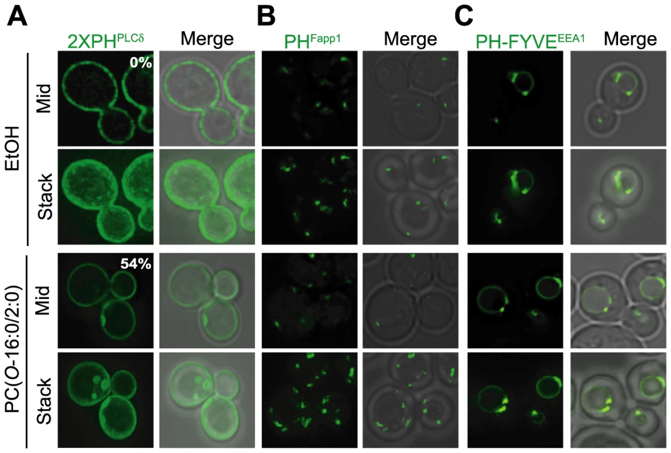
We had previously shown that PC(O-16 : 0/2 : 0) exposure led to the redistribution of the yeast PLD Spo14 at the PM into discrete foci [9]. As PLD activity is required to buffer the toxic effects of this lipid in both budding yeast and murine N2A neuroblastoma cells [9], we sought to discern the mechanism underlying the changes in PLD distribution. Since the localization of this enzyme to the PM is dependent upon interactions with PtdIns(4,5)P2, we examined the effects of PC(O-16 : 0/2 : 0) on the distribution of this lipid using a fluorescent probe for PtdIns(4,5)P2, GFP-2×PHPLCδ (Fig. 1A) [12], [20]–[22]. Similarly to Spo14, growth in the presence of PC(O-16 : 0/2 : 0) resulted in the relocalization of the GFP-tagged reporter construct to distinct membrane associated structures at the PM which we have termed PtdIns(4,5)P2 enriched structures (PES) (Fig. 1A). The appearance of the PES was maximal after 15 min of treatment with PC(O-16 : 0/2 : 0) and persisted for up to 90 min (Figure S1). This result was specific for PC(O-16 : 0/2 : 0) as all other related lipids, chemicals and conditions examined did not result in PES formation (Table S1). Furthermore, the distribution of GFP-tagged probes with specificity for additional intracellular phosphoinositides, PtdIns4P (PHFapp1) and PtdIns3P (PH-FYVEEEA1), were unaltered by PC(O-16 : 0/2 : 0) treatment suggesting a specific effect of this lipid on PM PtdIns(4,5)P2 (Fig. 1B and C) [20], [23].
PC(O-16 : 0/2 : 0) disrupts PM MSS4 distribution
The abundance of PtdIns(4,5)P2 depends upon the opposing actions of Mss4 and multiple PtdIns(4,5)P2 phosphatases including Inp51, Inp52 and Inp54 (reviewed in [24]). Similar to our previous findings with GFP-tagged Spo14 and GFP-2×PHPLCδ, PC(O-16 : 0/2 : 0) treatment resulted in the relocalization of Mss4-GFP to distinct foci within the cell (Fig. 2A). This result suggested that PC(O-16 : 0/2 : 0)-induced PES formation requires Mss4 activity. To investigate this possibility, we assessed PC(O-16 : 0/2 : 0)-induced PES formation in wild type cells and those carrying a thermosensitive allele of MSS4 (mss4-102) [20]. The reduced levels of PtdIns(4,5)P2 in mss4-102 cells precluded the use of GFP-2×PHPLCδ [20]–[22]. Therefore, changes in the PM structure were visualized using the lipophillic probe FM4-64, which co-localizes with GFP-2×PHPLCδ following PC(O-16 : 0/2 : 0) treatment in wild type cells (Fig. S2). As expected, both wild type and mss4-102 cells grown at the permissive temperature (25 C) exhibit similar FM4-64 labeling that was restricted to the PM and early endosomes in untreated cells (Fig. 2B). Following treatment with PC(O-16 : 0/2 : 0), structures similar to the PES were observed to form in both strains (Fig. 2B). Growth at the restrictive temperature did not impact PES formation in wild type cells as the formation of these structures was similar to previous results with maximal PES formation evident at 15 min and persisting for at least 60 min (Fig. 2B and Fig. S2E). However, PES formation was reduced in mss4-102 cells at all examined time points (Fig. 2B and Fig. S2E) suggesting that Mss4 activity is involved in PES formation. To assess the significance of Mss4-dependent PtdIns(4,5)P2 synthesis in buffering against PC(O-16 : 0/2 : 0) toxicity, we examined the growth of strains possessing temperature sensitive alleles of MSS4 (i.e. mss4-102) and the PtdIns 4-kinase STT4 (i.e. stt4-4) [20], [25]. Both mutant strains displayed increased sensitivity to PC(O-16 : 0/2 : 0) compared to the isogenic wild type control whereas overexpressing Mss4 reduced the growth inhibitory effects of PC(O-16 : 0/2 : 0) in an otherwise wild type strain (Fig. 2C and Fig. S2F). Furthermore, growth was also impacted by reducing or increasing the cellular PtdIns(4,5)P2 levels through overexpressing or deleting phosphoinositide phosphatases respectively (Fig. 2D and Fig. S2G–H). In particular, overexpression of Inp51 and Inp54 resulted in reduced growth whereas deletion of Inp51 alone improved growth in the presence of PC(O-16 : 0/2 : 0) (Fig. 2D and Fig. S2G) [24]. Together these results indicate that cellular PtdIns(4,5)P2 and PES formation are important for buffering against the toxic effects of PC(O-16 : 0/2 : 0).

The PES are PM invaginations that form independently of the actin cytoskeleton
We next sought to investigate the cellular processes involved in PES formation. First, we examined the ultrastructure of the PES by electron microscopy (EM). In contrast to those untreated, cells exposed to PC(O-16 : 0/2 : 0) displayed large invaginations of the PM, which occasionally appeared as either a transversal cut of the PM invagination or potentially invaginations which have undergone scission and become cytoplasmic (Fig. 3A–F and Fig. S3A). The large invaginations of the PM present in PC(O-16 : 0/2 : 0) treated cells are reminiscent of the failed endocytic events that have previously been observed in inp51Δ inp52Δ cells [26]–[28]. The formation of these structures in the inp51Δ inp52Δ mutant is due to increased PtdIns(4,5)P2 levels as a result of reduced cellular PtdIns(4)P 5-phosphatase activity [28]. This phenomenon requires an intact actin cytoskeleton [28]. In contrast, pretreatment with Latrunculin A (Lat A), an actin depolymerizing agent, did not inhibit PES formation (Fig. 3G) and surprisingly we found that PC(O-16 : 0/2 : 0) treatment alone resulted in the disruption of the actin cytoskeleton (Fig. 3H). Similarly, deletion of VRP1, an actin associated protein required for cytoskeletal organization that suppresses the inp51Δ inp52Δ phenotype [29], did not affect PES formation or PC(O-16 : 0/2 : 0) toxicity (Fig. S3B and C). Combined these results strongly suggest that the PC(O-16 : 0/2 : 0)-dependent PES is distinct from the previously characterized PM invaginations seen in inp51Δ inp52Δ cells and that the PES formation occurs independently of the actin cytoskeleton. The actin-independency of PES formation could potentially be explained by an unregulated association of endocytic coat complex proteins or impaired exocytic vesicle fusion [30]. However, a RFP-fusion of Chc1, which associates at the PM independently of actin at sites of clathrin-mediated endocytosis [31], co-localized with GFP-2×PHPLCδ at the PES in only 3% of cells (Fig. S3D). In addition, the localization of the exocyst component Exo70 was only modestly disrupted upon PC(O-16 : 0/2 : 0) treatment (Fig. S3E) and both Exo70-GFP or Sec3-GFP exhibited minimal co-localization with the PES marked by FM4-64 (Fig. S3F). These results indicate that the actin-independent events involved in PES formation likely do not involve the aberrant association of endocytic or exocytic proteins with the PM.

PC(O-16 : 0/2 : 0) disrupts sphingolipid metabolism
These findings suggested that Mss4 relocalization is a principal factor in PES formation and that perturbations to PM PtdIns(4,5)P2 distribution are critically involved in regulating the toxic effects of PC(O-16 : 0/2 : 0). How might PC(O-16 : 0/2 : 0) disrupt Mss4 localization? The association of this protein with the PM occurs through poorly defined processes and may involve a combination of protein-protein and lipid-protein interactions [30], [32], [33]. Interestingly, the only reported lipid factors mediating Mss4 localization to the PM are PtdIns(4)P and the complex sphingolipid mannose-inositol-phosphoceramide (MIPC) [30], [33]. Although the role of MIPC was not confirmed by a subsequent study [34], Gallego and co-workers have shown that Mss4 can bind to dihydrosphingosine-1 phosphate (DHS-1P) in vitro and that an extended treatment with an inhibitor of sphingolipid biosynthesis (myriocin, 2 h) results in relocalization of Mss4-GFP [32]. These results suggest that changes in sphingolipid levels can impact Mss4 localization. Therefore, we postulated that the biological consequences of PC(O-16 : 0/2 : 0) treatment may arise in response to the effects of PC(O-16 : 0/2 : 0) on either sphingolipid biosynthesis or catabolism. In agreement with this hypothesis, we observed a global accumulation of LCBs precursors, their phosphorylated derivatives (LCB-Ps), as well as immediate ceramide precursors and metabolites in cells treated with PC(O-16 : 0/2 : 0) for 90 min (Fig. 4A, Dataset 1 and Fig. S4B). Furthermore, a modest but significant increase in several unphosphorylated phytosphingosine (PHS) and dihydrosphingosine (DHS) species is evident at 15 min (Fig. S4B). We also report that these increases were not associated with a decrease in the abundance of complex sphingolipids suggesting that PC(O-16 : 0/2 : 0) does not induce their catabolism (Fig. 4A, Fig. S4D and Dataset S1). In addition, deletion of the S. cerevisiae enzyme required for catabolism of complex sphingolipids, ISC1, did not impact the effects of PC(O-16 : 0/2 : 0) upon cell growth, PES formation or sphingolipid levels indicating that PC(O-16 : 0/2 : 0) does not stimulate the breakdown of sphingolipids (Fig. S4B–D). Next, we sought to determine whether PC(O-16 : 0/2 : 0)-induced elevation in LCBs and/or ceramide levels contributed to PES formation. First, we directly assessed the effects of ceramide upon PES formation by treating cells with the cell permeable ceramide, Cer(d18 : 1/2 : 0), or a biologically inactive analog, Cer(d18 : 0/2 : 0) (Fig. 4B). Treatment with Cer(d18 : 1/2 : 0), but not Cer(d18 : 0/2 : 0) resulted in relocalization of PtdIns(4,5)P2 and depolarization of the actin cytoskeleton similar to what is observed upon exposure to PC(O-16 : 0/2 : 0) suggesting that elevated ceramide levels are sufficient to induce PES formation (Fig. 4B). To explore the role of PC(O-16 : 0/2 : 0)-induced accumulation of LCB and ceramide further, we next investigated the effects of myriocin, an inhibitor of sphingolipid biosynthesis [35] (Fig. S4A), on Mss4-GFP localization in PC(O-16 : 0/2 : 0) treated cells (Fig. 4C and S4A). To accomplish this, we first pretreated cells with myriocin for 30 minutes prior to exposing them to PC(O-16 : 0/2 : 0). Although longer exposure (2 h) to myriocin has been reported to impact Mss4-GFP localization [32], our short pretreatment with myriocin did not affect Mss4-GFP localization (Fig. 4C). Pretreatment with myriocin for this time period was sufficient to inhibit the relocalization of Mss4-GFP and PES formation induced by PC(O-16 : 0/2 : 0) (Fig. 4C and Fig. S4F). Combined, these results support the notion that PC(O-16 : 0/2 : 0) treatment promotes the accumulation of LCBs and ceramides, which in turn contribute to changes in the subcellular localization of Mss4-GFP, PtdIns(4,5)P2 and downstream signaling events including actin cytoskeleton polarization.

PC(O-16 : 0/2 : 0) inhibits Tor2 signaling
We next sought to identify relevant signaling pathways which might be impacted by the effects of PC(O-16 : 0/2 : 0) upon sphingolipid metabolism and PM PtdIns(4,5)P2 localization. The target of rapamycin complex 2 (TORC2) was identified as a potential target because of its localization to the PM and the responsiveness of this signaling complex to changes in sphingolipid biosynthesis [36]–[38]. Furthermore, TORC2 has an established role in maintaining actin cytoskeleton polarization which is dependent upon the PM recruitment and phosphorylation of the homologous kinases Ypk1 and Ypk2 by the PtdIns(4,5)P2 binding proteins Slm1 and Slm2 [39], [40]. Utilizing a phospho-specific antibody recognizing a TORC2-dependent phosphorylation site on Ypk1 (T662) we determined that phosphorylation of endogenous Ypk1 was reduced in PC(O-16 : 0/2 : 0) suggesting that TORC2 signaling is inhibited by PC(O-16 : 0/2 : 0) (Fig. 5A) [37].

A critical role for Tor2 and Ypk kinase signaling in PC(O-16 : 0/2 : 0) toxicity
Similar to mammalian cells, two distinct multiprotein complexes containing Tor activity, i.e. TORC1 and TORC2, are present in yeast. Unlike mammalian cells, however, yeast possess two TOR genes, TOR1 and TOR2, with Tor1 nucleating the formation of TORC1 while Tor2 is able to nucleate both TORC1 and TORC2 [41]. Given that the phosphorylation of the TORC2 target Ypk1 is potently inhibited by PC(O-16 : 0/2 : 0), we next sought to determine whether Tor2 activity is required for preventing the growth inhibitory effects of PC(O-16 : 0/2 : 0). To assess the relative role of each Tor protein in buffering the growth inhibitory effects of PC(O-16 : 0/2 : 0), we made use of strains harboring the temperature sensitive tor2-21 and tor2-30 alleles alone or in combination with deletion of TOR1 [42]. Whereas deletion of TOR1 alone had no observable effect upon PC(O-16 : 0/2 : 0) sensitivity (Fig. 5B), the tor2-21 strain exhibited a significant reduction in growth in the presence of PC(O-16 : 0/2 : 0) at a semi-permissive temperature. To further validate the role of TORC2 signaling in mediating PC(O-16 : 0/2 : 0) sensitivity we examined the effect of overexpressing the downstream target YPK2 [40]. Consistent with a role for TORC2 in mediating the response to PC(O-16 : 0/2 : 0), we found that overexpression of a YPK2 hyperactive allele (D239A), known to rescue lethality of TORC2 mutants [40], was able to restore growth of the tor2-21 strain in the presence of PC(O-16 : 0/2 : 0). Comparatively, the wild type (Ypk2) and the kinase dead (K373A) variants [40] were unable to restore growth in the presence of reduced Tor2 function (Fig. 5C). Together, these results provide compelling evidence that TORC2 is inhibited in response to PC(O-16 : 0/2 : 0) treatment and that a reduction in TORC2 signaling is associated with an increased sensitivity to PC(O-16 : 0/2 : 0).
Examining the mechanism underlying Tor2 inhibition by PC(O-16 : 0/2 : 0)
Since these results establish an important role for the TORC2-Ypk2 signaling in mediating the cellular response to PC(O-16 : 0/2 : 0), we investigated the potential mechanisms by which PC(O-16 : 0/2 : 0) might act to inhibit TORC2-dependent phosphorylation of Ypk1/2. The requirement for PtdIns(4,5)P2, PLD and Tor signaling in mediating PC(O-16 : 0/2 : 0) sensitivity presented the intriguing possibility that PLD-generated PA regulates Tor signaling in S. cerevisiae as previously reported for mTor [43]–[46]. However, deletion of SPO14, did not have noticeable effected the phosphorylation of endogenous Ypk1 suggesting that Spo14 does not impact TORC2 function in S. cerevisiae (Fig. 5A). Furthermore, knock out of SPO14 exhibited a synthetic interaction with the tor2-21 allele (Fig. S5). These results indicate that Spo14 and Tor2 likely act through parallel signaling pathways. Alternatively, the inhibition of Ypk1 phosphorylation in PC(O-16 : 0/2 : 0)-treated cells may be due to the direct inhibition of Tor kinase activity as was previously reported for cells with elevated glycerophosphocholine levels [47]. PC(O-16 : 0/2 : 0), however, did not inhibit the phosphorylation of recombinant GST-Ypk2 by immunopurified TORC2 suggesting PC(O-16 : 0/2 : 0) does not act as a direct inhibitor of Tor function in vitro and that a secondary mediator is required (Fig. S6A). Given that Ypk1/2 and TORC2 are normally localized to distinct subcellular compartments, however, the in vitro kinase assay likely does not fully recapitulate the constraints present in vivo. For example, phosphorylation of Ypk1/2 requires relocalization from the cytosol to the PM by TORC2 adaptor proteins Slm1/2 [48]. Interestingly, localization of Slm1/2 at the PM is itself partly dependent upon interactions with PtdIns(4,5)P2 [21], [34], [48]. We observed that PC(O-16 : 0/2 : 0) treatment disrupted the typical association of Slm1-GFP with eisosomes, a distinct spatially segregated compartment of the PM in S. cerevisiae [49], as indicated by the reduction in co-localization of Slm1-GFP with a tagged eisosome protein, Lsp1-mCherry (Fig. 6A). This redistribution of Slm1-GFP was not due to disruption of eisosome integrity but was associated with its appearance at the PES (Fig. S6B–D). Furthermore overexpression of Slm1 from a high copy plasmid enhanced growth compared to vector alone suggesting that Slm1-dependent signaling events are critically involved in mediating the cellular response to PC(O-16 : 0/2 : 0) (Fig. S6E). The correlation of Slm1 relocalization with increased LCBs and ceramides (Fig. 4 and S4) in PC(O-16 : 0/2 : 0)-treated cells is complementary with a previous report describing the impact of inhibiting sphingolpid metabolism upon the subcellular localization of Slm1 and Ypk1 phosphorylation [37]. Therefore, we next sought to investigate whether the relocalization of Slm1-GFP in PC(O-16 : 0/2 : 0) impaired the interaction of Ypk1 or TORC2. However, we found that the association of Slm1-GFP with HA-tagged TORC2 component Avo3 or untagged Ypk1 was not affected by PC(O-16 : 0/2 : 0) treatment suggesting the inhibition of TORC2 signaling does not require the redistribution of Slm1 to the PES (Fig. 6B). To support this conclusion we next investigated whether PES formation was necessary for the PC(O-16 : 0/2 : 0)-dependent inhibition of Ypk1 phosphorylation (Fig. 6C). Although pretreatment with myriocin alone increased Ypk1 phosphorylation ∼2.3 fold we observed that phosphorylation was similarly reduced (∼50%) in cells pretreated with either vehicle or myriocin upon treatment with PC(O-16 : 0/2 : 0) (Fig. 6C). Therefore, the inhibition of TORC2-dependent Ypk1 phosphorylation by PC(O-16 : 0/2 : 0) likely does not require the recruitment of Slm1 to the PES or a reduced interaction of Ypk1 with Slm1 or Avo3, indicating PC(O-16 : 0/2 : 0) inhibiting TORC2 through an previously undescribed mechanism.

Discussion
Aberrant glycerophosphocholine metabolism in AD leading to the intraneuronal accumulation of specific lipid second messengers, including PC(O-16 : 0/2 : 0) is linked to neuronal dysfunction, neurotoxicity, and accelerated cognitive decline [6], [50]–[52]. In this report we have used S. cerevisiae to further characterize the mechanisms underlying receptor-independent toxicity of PC(O-16 : 0/2 : 0). Our work suggests a model (Fig. 7) wherein exposure to toxic concentrations of PC(O-16 : 0/2 : 0) promotes the accumulation of LCBs and ceramides, which leads to changes in the subcellular localization of Mss4 and formation of PtdIns(4,5)P2 enriched invaginations of the PM. Ultimately the PC(O-16 : 0/2 : 0)-dependent remodeling of PtdIns(4,5)P2 affects downstream PtdIns(4,5)P2-dependent cellular processes such as PLD localization, which is critical for buffering against the toxic effects of PC(O-16 : 0/2 : 0) [9]. However, the inhibition of TORC2 by PC(O-16 : 0/2 : 0) also suggests that the toxic properties of PC(O-16 : 0/2 : 0) are only partly due to disruptions in PtdIns(4,5)P2 signaling and that this lipid impacts other signaling pathways through distinct second messengers that remain to be identified.

Given that PtdIns(4,5)P2 and downstream signaling events buffer against PC(O-16 : 0/2 : 0) toxicity, it was important to investigate the factors underlying the relocalization of Mss4-GFP and PES formation to elucidate potential endogenous mechanisms of neuroprotection. The molecular details that contribute to the localization of Mss4 into distinct phosphatidylinositol kinase or PIK patches in yeast are not completely understood. However, the availability of its substrate, PtdIns(4)P, a recently identified interacting partner Opy1 and sphingolipid biosynthesis have been implicated [30], [32], [33]. Our data suggests that the PC(O-16 : 0/2 : 0)-induced accumulation of LCBs and ceramides (precursor molecules in the sphingolipid biosynthetic pathway Fig. S4) are at least partly responsible for the changes in PM PtdIns(4,5)P2 distribution as treatment with myriocin, an inhibitor of sphingolipid biosynthesis, was sufficient to prevent the redistribution of Mss4-GFP and PES formation (Fig. 4 and Fig. S4). The mechanism by which the observed changes in LCBs and ceramide might regulate Mss4 PM localization are not clear but previous reports have suggested that both MIPC and dihydrosphingosine-1 phosphate (DHS-1P) can interact with Mss4 [32], [33]. The relocalization of Mss4-GFP, however, is likely not due to interactions with MIPC as neither the total levels of this lipid nor the abundance of individual species was significantly impacted by PC(O-16 : 0/2 : 0) at any time point (Fig. 4, Fig. S4 and Dataset 1). In contrast, the accumulation of one DHS and two PHS species displayed similar kinetics to the PtdIns(4,5)P2 redistribution and PES formation suggesting that these lipids may be involved in mediating the observed changes (Fig. S4B and Dataset 1). Certainly, this observation must be interpreted with caution as the reported in vitro interaction between Mss4 and LCBs has not been evaluated in vivo [32]. Furthermore, the role of LCB-Ps in mediating Mss4 localization at the PM must also be reconciled with the fact that LCB-Ps do not appear to be trafficked to the PM under normal circumstances [53]. Whether PC(O-16 : 0/2 : 0)-induced changes in Mss4-GFP localization are dependent upon the improper trafficking of LCB-Ps or another mechanism remains an open question in need of further study.
The spatial distribution of PtdIns(4,5)P2 at the PM is critical for regulating the activity of downstream signaling pathways. Our biochemical, genetic and cell biology based-assays suggest that the inhibition of Tor signaling plays a critical role in mediating the sensitivity to the toxic effects of PC(O-16 : 0/2 : 0) (Fig. 5 and 6). The results of our in vitro kinase assay do not identify PC(O-16 : 0/2 : 0) as a direct inhibitor of Tor kinase activity and suggests that another mechanism is responsible for the inhibition of Tor signaling (Fig. S6). How else might PC(O-16 : 0/2 : 0) inhibit Tor signaling? Although the cellular inputs which impinge upon Tor signaling are still being identified and the molecular mechanisms which translate these stimuli into activation/inhibition of Tor signaling are not completely understood, the TORC2-dependent phosphorylation of Ypk1/2 is sensitive to changes in PM PtdIns(4,5)P2 levels [48]. Our work demonstrating the relocalization of Slm1-GFP to the PES in response to PC(O-16 : 0/2 : 0) is consistent with previous reports describing the interactions of Slm proteins with PtdIns(4,5)P2 (Fig. 6 and S6) [1], [20]. Because our data indicate that relocalization of Slm1, and presumably Slm2, to the PES is not required for the inhibition of TORC2-dependent Ypk phosphorylation, they suggest that an additional mechanism(s) exists for the regulation of TORC2 signaling (Fig. 6).
Collectively, our results provide insight into how a disruption in phosphocholine metabolism signals network-wide lipid metabolic disturbances that may play defining roles into how neurons respond to accumulating Aβ42. Interestingly, accumulating evidence suggests that disruptions in both PtdIns(4,5)P2 signaling and ceramide metabolism are contributing factors in the neuronal cell dysfunction and death observed in AD [13]–[19]. Whether the disruptions in PtdIns(4,5)P2 signaling and ceramide metabolism homeostasis observed in neurons are dependent upon an increase in PC(O-16 : 0/2 : 0) concentrations is an intriguing question in need of further investigation.
Materials and Methods
Yeast strains, plasmids and media
The yeast strains and plasmids used in this study are listed in Table S2 and S3. Strains were generated by using a standard PCR-mediated gene insertion/deletion technique [54]. Cells were grown in standard YPD or SD medium supplemented with amino acids and all lipids were prepared by resuspending in either ethanol or methanol and storing under nitrogen gas.
Cell growth and treatments
All strains were grown in YPD or minimal media supplemented with appropriate amino acids as required and treated with PC(O-16 : 0/2 : 0) (Enzo Life Sciences, BML-L100 or Avanti Polar Lipids, 878119P) at 20 µM for 15 minutes unless indicated otherwise. Media was supplemented with rapamycin (200 ng/ml) where indicated.
Dot assays
Cells were grown in YPD or minimal media at 30 C to mid-log phase and resuspended to an OD600 of 0.1. Dot assays were performed by spotting 4 µL of ten-fold serial dilutions (OD600 = 0.1, 0.01, 0.001, 0.0001) onto YPD or minimal media selection plates containing the specified concentrations of ethanol, PC(O-16 : 0/2 : 0) or other chemical as indicated.
Microscopy
For all microscopy experiments, overnight cultures grown at 30 C in YPD medium were re-suspended at a final OD600 of 0.2 and allowed to reach mid-log phase prior treatment and image acquisition. Live cell imaging was performed by briefly centrifuging the cells (800 g for 3 min), followed by resuspending in a minimal volume of growth media, spotting onto glass slides and coverslipping prior to imaging. All images were acquired using a Leica DMI 6000 florescent microscope (Leica Microsystems GmbH, Wetzler Germany), equipped with a Sutter DG4 light source (Sutter Instruments, California, USA), Ludl emission filter wheel with Chroma band pass emission filters (Ludl Electronic Products Ltd., NY, USA) and Hamamatsu Orca AG camera (Hamamatsu Photonics, Herrsching am Ammersee, Germany). Images were acquired at 0.2 µM steps using a 63× oil-immersion objective with a 1.4 numerical aperture. Deconvolution and analysis were performed using Velocity Software V4 (Perkin Elmer). For most images, representative images of the middle section and compressed image stack are shown. Numerical insets represent the indicated quantifications of at least 100 cells from 2 to 3 independent experiments unless indicated otherwise.
Rhodamine-phalloidin staining
Early log phase cells were fixed by diluting 37% formaldehyde to a final concentration of 3.7% and incubating at 25 C for 10 minutes. Cells were subsequently pelleted (800 g for 3 min) and resuspended in PBS containing 3.7% formaldehyde and incubated for 1 hour. Cells were subsequently washed three times in PBS prior to staining with Rhodamine-conjugated phalloidin diluted in PBS containing 0.1% Tween on ice (20 Units/ml, Invitrogen) and cells were washed two times prior to imaging. For actin depolymerisation, Latrunculin A (5 µM, Tocris) was added as indicated prior to fixation and cell staining.
Cell extract preparation
In all cases overnight cultures of yeast strains were diluted to an OD600 of 0.2 in YPD or appropriate minimal media and allowed to reach mid-log growth prior to harvesting. Cell pellets were resuspended in 200 µL of lysis buffer (20 mM HEPES, 150 mM NaCl, 2 mM EDTA with phosphatase and protease inhibitors and lysed by vortexing with glass beads. Ypk1 was examined in ethanol and PC(O-16 : 0/2 : 0) treated cells prepared as previously described [37]. Briefly, ice cold acetone was added to mid log phase cells and incubate on ice for 5 min. Cells were pelleted and washed two times in 5% acetone in PBS. Supernatant was removed and the cells pellets were dried under vacuum prior to lysis in urea buffer.
Electron microscopy
Processing for electron microscopy was performed as previously described [55].
TORC2 in vitro kinase assay
TORC2 was purified from RL127-1c cells. The cultures were grown to an OD600 of 5.0 in YPD (125 mL per assay point), chilled on ice for 30 minutes, collected, and washed. The cells were put into liquid nitrogen and ground up using a mortar and pestle. The powder was then resuspended in lysis buffer (1× Roche protease inhibitor +EDTA, 1 mM PMSF, phosphatase inhibitors, 5 mM CHAPS, 50 mM HEPES pH 7.5, 300 mM KCl), spun down, and 420 ul of prepared paramagnetic beads (Dynabeads M-270 Epoxy, coated with rabbit IgG; Sigma) were added to the cleared protein extracts. The tubes were subsequently rotated for 3 h at 4°C. Beads were collected by using a magnet and washed extensively with lysis buffer. The kinase reactions were performed in a final volume of 30 µl containing TORC2-coupled beads, 300 ng of Ypk2, 25 mM Hepes pH 7.0, 50 mM KCl, 4 mM MgCl2, 10 mM DTT, 0.5% Tween20, 1× Roche protease inhibitor-EDTA, 100 mM ATP, 5 mCi [γ-32P]-ATP and 1 µl of inhibitors at various concentrations. PAF was dissolved in EtOH and used at the indicated concentrations. Assays were started with addition of ATP, maintained at 30°C for 25 minutes and terminated by the addition of 7.5 µl of 5× SDS-PAGE buffer. Samples were heated at 65°C for 10 min; proteins were resolved in SDS-PAGE, stained with Sypro Ruby and analysed using a Bio-Rad Molecular Imager.
Substrate preparation for in vitro kinase assays
GST-Ypk2 fusion proteins were expressed in S. cerevisiae from a pRS426 vector. Actively growing cells were induced for 3 hours with galactose (final concentration of 2%), chilled on ice for 30 minutes, and collected. The cells were put into liquid nitrogen and ground up using a mortar and pestle. The powder was then resuspended in lysis buffer (10% glycerol, 1×PBS, 0.5% Tween, 1× Roche protease inhibitor +EDTA, 1 mM PMSF, and phosphatase inhibitors) and the fusion protein was bound to and eluted from glutathione Sepharose 4B (GE Healthcare) following standard procedures. The supernatant was dialyzed against ( 50% glycerol, 1 mM DTT, 1 mM EDTA, 25 mM Tris pH 7.5, 50 mM NaCl), aliquotted, and frozen at −20°C.
PC(O-16 : 0/2 : 0) treatment and lipid extraction
Cells at 0.6 OD600 were treated with 20 µM PAF or ethanol as a control. At T = 15 min, 30 min , 60 min and 120 min, 7.5 OD600 were harvested in glass tubes, washed with water and the pellet was extracted 3×1 ml in water∶Ethanol∶Diethyl Ether∶Pyridine∶NH4OH (15∶15∶5∶1∶0.018) at 65°C for 15 min each time. Avanti Polar Lipid MS standards (LM-6002) were added during the first extraction at 62.5 pmol/tube. The extracts were pooled and dried under N2, redissolved in 1 ml Chloroform with bath sonication, 1 ml Butanol was added and phospholipids were hydrolyzed for 30 min at 37°C after the addition of 200 µL 1 M KOH (in methanol). After hydrolysis, the extract was neutralized by the addition of 200 µL 1 M Acetic Acid (in Methanol). 1 ml Butanol saturated water was added, centrifuged to separate the phases and the upper aqueous layer was removed by aspiration, being careful not to disrupt the precipitate at the interface. This was repeated two more times after which the remaining lower phase was dried under N2. The dried lipid was redissolved in 0.5 ml LC/MS buffer A with bath sonication, spun to pellet insoluble material and the transferred to MS analysis vials.
LC/MS analysis
The samples were analyzed on a Supelco Discovery Bio Wide Pore C18 (5 cm×2.1 mm, 5 uM) column at 40°C (50 mm) using an Agilent 1200 Series HPLC coupled to ABSciex QTRAP 4000 MS. The LCB(P)s were eluted using a binary solvent gradient of 0% B for 1 min, 25% at 4 min, 100% at 4.5 min and held at 100%B for 1.5 min, 0%B at 7 min. The LCB(P)s were detected in MRM mode,
LC/MS buffers
MS buffer A: Tetrahydrofuran: Methanol: 10 mM Ammonium Formate (30∶20∶50) with 0.2% Formic Acid
MS buffer B: Tetrahydrofuran: Methanol: 10 mM Ammonium Formate (70∶20∶10) with 0.2% Formic Acid
Supporting Information
Zdroje
1. WymannMP, SchneiterR (2008) Lipid signalling in disease. Nat Rev Mol Cell Biol 9 : 162–176.
2. Di PaoloG, KimTW (2011) Linking lipids to Alzheimer's disease: cholesterol and beyond. Nat Rev Neurosci 12 : 284–296.
3. WoodPL (2012) Lipidomics of Alzheimer's disease: current status. Alzheimers Res Ther 4 : 5.
4. ChanRB, OliveiraTG, CortesEP, HonigLS, DuffKE, et al. (2012) Comparative lipidomic analysis of mouse and human brain with Alzheimer disease. J Biol Chem 287 : 2678–2688.
5. RyanSD, HarrisCS, MoF, LeeH, HouST, et al. (2007) Platelet activating factor-induced neuronal apoptosis is initiated independently of its G-protein coupled PAF receptor and is inhibited by the benzoate orsellinic acid. J Neurochem 103 : 88–97.
6. RyanSD, WhiteheadSN, SwayneLA, MoffatTC, HouW, et al. (2009) Amyloid-beta42 signals tau hyperphosphorylation and compromises neuronal viability by disrupting alkylacylglycerophosphocholine metabolism. Proc Natl Acad Sci U S A 106 : 20936–20941.
7. RyanSD, HarrisCS, CarswellCL, BaenzigerJE, BennettSA (2008) Heterogeneity in the sn-1 carbon chain of platelet-activating factor glycerophospholipids determines pro - or anti-apoptotic signaling in primary neurons. J Lipid Res 49 : 2250–2258.
8. SantosAX, RiezmanH (2012) Yeast as a model system for studying lipid homeostasis and function. FEBS Lett 586 : 2858–2867.
9. KennedyMA, KabbaniN, LambertJP, SwayneLA, AhmedF, et al. (2011) Srf1 is a novel regulator of phospholipase D activity and is essential to buffer the toxic effects of C16 : 0 platelet activating factor. PLoS Genet 7: e1001299.
10. RudgeSA, MorrisAJ, EngebrechtJ (1998) Relocalization of phospholipase D activity mediates membrane formation during meiosis. J Cell Biol 140 : 81–90.
11. SciorraVA, RudgeSA, PrestwichGD, FrohmanMA, EngebrechtJ, et al. (1999) Identification of a phosphoinositide binding motif that mediates activation of mammalian and yeast phospholipase D isoenzymes. EMBO J 18 : 5911–5921.
12. SciorraVA, RudgeSA, WangJ, McLaughlinS, EngebrechtJ, et al. (2002) Dual role for phosphoinositides in regulation of yeast and mammalian phospholipase D enzymes. J Cell Biol 159 : 1039–1049.
13. LandmanN, JeongSY, ShinSY, VoronovSV, SerbanG, et al. (2006) Presenilin mutations linked to familial Alzheimer's disease cause an imbalance in phosphatidylinositol 4,5-bisphosphate metabolism. Proc Natl Acad Sci U S A 103 : 19524–19529.
14. BermanDE, Dall'ArmiC, VoronovSV, McIntireLB, ZhangH, et al. (2008) Oligomeric amyloid-beta peptide disrupts phosphatidylinositol-4,5-bisphosphate metabolism. Nat Neurosci 11 : 547–554.
15. McIntireLB, BermanDE, MyaengJ, StaniszewskiA, ArancioO, et al. (2012) Reduction of synaptojanin 1 ameliorates synaptic and behavioral impairments in a mouse model of Alzheimer's disease. J Neurosci 32 : 15271–15276.
16. FilippovV, SongMA, ZhangK, VintersHV, TungS, et al. (2012) Increased ceramide in brains with Alzheimer's and other neurodegenerative diseases. J Alzheimers Dis 29 : 537–547.
17. HanX, RozenS, BoyleSH, HellegersC, ChengH, et al. (2011) Metabolomics in early Alzheimer's disease: identification of altered plasma sphingolipidome using shotgun lipidomics. PLoS One 6: e21643.
18. HanX, DMH, McKeelDWJr, KelleyJ, MorrisJC (2002) Substantial sulfatide deficiency and ceramide elevation in very early Alzheimer's disease: potential role in disease pathogenesis. J Neurochem 82 : 809–818.
19. CutlerRG, KellyJ, StorieK, PedersenWA, TammaraA, et al. (2004) Involvement of oxidative stress-induced abnormalities in ceramide and cholesterol metabolism in brain aging and Alzheimer's disease. Proc Natl Acad Sci U S A 101 : 2070–2075.
20. StefanCJ, AudhyaA, EmrSD (2002) The yeast synaptojanin-like proteins control the cellular distribution of phosphatidylinositol (4,5)-bisphosphate. Mol Biol Cell 13 : 542–557.
21. FadriM, DaquinagA, WangS, XueT, KunzJ (2005) The pleckstrin homology domain proteins Slm1 and Slm2 are required for actin cytoskeleton organization in yeast and bind phosphatidylinositol-4,5-bisphosphate and TORC2. Mol Biol Cell 16 : 1883–1900.
22. AudhyaA, LoewithR, ParsonsAB, GaoL, TabuchiM, et al. (2004) Genome-wide lethality screen identifies new PI4,5P2 effectors that regulate the actin cytoskeleton. EMBO J 23 : 3747–3757.
23. BurdCG, EmrSD (1998) Phosphatidylinositol(3)-phosphate signaling mediated by specific binding to RING FYVE domains. Mol Cell 2 : 157–162.
24. StrahlT, ThornerJ (2007) Synthesis and function of membrane phosphoinositides in budding yeast, Saccharomyces cerevisiae. Biochim Biophys Acta 1771 : 353–404.
25. AudhyaA, FotiM, EmrSD (2000) Distinct roles for the yeast phosphatidylinositol 4-kinases, Stt4p and Pik1p, in secretion, cell growth, and organelle membrane dynamics. Mol Biol Cell 11 : 2673–2689.
26. StolzLE, HuynhCV, ThornerJ, YorkJD (1998) Identification and characterization of an essential family of inositol polyphosphate 5-phosphatases (INP51, INP52 and INP53 gene products) in the yeast Saccharomyces cerevisiae. Genetics 148 : 1715–1729.
27. Singer-KrugerB, NemotoY, DaniellL, Ferro-NovickS, De CamilliP (1998) Synaptojanin family members are implicated in endocytic membrane traffic in yeast. J Cell Sci 111(Pt 22): 3347–3356.
28. StefanCJ, PadillaSM, AudhyaA, EmrSD (2005) The phosphoinositide phosphatase Sjl2 is recruited to cortical actin patches in the control of vesicle formation and fission during endocytosis. Mol Cell Biol 25 : 2910–2923.
29. SunY, CarrollS, KaksonenM, ToshimaJY, DrubinDG (2007) PtdIns(4,5)P2 turnover is required for multiple stages during clathrin - and actin-dependent endocytic internalization. J Cell Biol 177 : 355–367.
30. LingY, StefanCJ, MacgurnJA, AudhyaA, EmrSD (2012) The dual PH domain protein Opy1 functions as a sensor and modulator of PtdIns(4,5)P(2) synthesis. EMBO J 31(13): 2882–94.
31. NewpherTM, SmithRP, LemmonV, LemmonSK (2005) In vivo dynamics of clathrin and its adaptor-dependent recruitment to the actin-based endocytic machinery in yeast. Dev Cell 9 : 87–98.
32. GallegoO, BettsMJ, Gvozdenovic-JeremicJ, MaedaK, MatetzkiC, et al. (2010) A systematic screen for protein-lipid interactions in Saccharomyces cerevisiae. Mol Syst Biol 6 : 430.
33. KobayashiT, TakematsuH, YamajiT, HiramotoS, KozutsumiY (2005) Disturbance of sphingolipid biosynthesis abrogates the signaling of Mss4, phosphatidylinositol-4-phosphate 5-kinase, in yeast. J Biol Chem 280 : 18087–18094.
34. TabuchiM, AudhyaA, ParsonsAB, BooneC, EmrSD (2006) The phosphatidylinositol 4,5-biphosphate and TORC2 binding proteins Slm1 and Slm2 function in sphingolipid regulation. Mol Cell Biol 26 : 5861–5875.
35. MiyakeY, KozutsumiY, NakamuraS, FujitaT, KawasakiT (1995) Serine palmitoyltransferase is the primary target of a sphingosine-like immunosuppressant, ISP-1/myriocin. Biochem Biophys Res Commun 211 : 396–403.
36. BerchtoldD, WaltherTC (2009) TORC2 plasma membrane localization is essential for cell viability and restricted to a distinct domain. Mol Biol Cell 20 : 1565–1575.
37. BerchtoldD, PiccolisM, ChiaruttiniN, RiezmanI, RiezmanH, et al. (2012) Plasma membrane stress induces relocalization of Slm proteins and activation of TORC2 to promote sphingolipid synthesis. Nat Cell Biol 14 : 542–547.
38. AronovaS, WedamanK, AronovPA, FontesK, RamosK, et al. (2008) Regulation of ceramide biosynthesis by TOR complex 2. Cell Metab 7 : 148–158.
39. SchmidtA, KunzJ, HallMN (1996) TOR2 is required for organization of the actin cytoskeleton in yeast. Proc Natl Acad Sci U S A 93 : 13780–13785.
40. KamadaY, FujiokaY, SuzukiNN, InagakiF, WullschlegerS, et al. (2005) Tor2 directly phosphorylates the AGC kinase Ypk2 to regulate actin polarization. Mol Cell Biol 25 : 7239–7248.
41. LoewithR, HallMN (2011) Target of rapamycin (TOR) in nutrient signaling and growth control. Genetics 189 : 1177–1201.
42. HelliwellSB, HowaldI, BarbetN, HallMN (1998) TOR2 is part of two related signaling pathways coordinating cell growth in Saccharomyces cerevisiae. Genetics 148 : 99–112.
43. FangY, ParkIH, WuAL, DuG, HuangP, et al. (2003) PLD1 regulates mTOR signaling and mediates Cdc42 activation of S6K1. Curr Biol 13 : 2037–2044.
44. FangY, Vilella-BachM, BachmannR, FlaniganA, ChenJ (2001) Phosphatidic acid-mediated mitogenic activation of mTOR signaling. Science 294 : 1942–1945.
45. YoonMS, SunY, ArauzE, JiangY, ChenJ (2011) Phosphatidic acid activates mammalian target of rapamycin complex 1 (mTORC1) kinase by displacing FK506 binding protein 38 (FKBP38) and exerting an allosteric effect. J Biol Chem 286 : 29568–29574.
46. ToschiA, LeeE, XuL, GarciaA, GadirN, et al. (2009) Regulation of mTORC1 and mTORC2 complex assembly by phosphatidic acid: competition with rapamycin. Mol Cell Biol 29 : 1411–1420.
47. ZhangC, WendelAA, KeoghMR, HarrisTE, ChenJ, et al. (2012) Glycerolipid signals alter mTOR complex 2 (mTORC2) to diminish insulin signaling. Proc Natl Acad Sci U S A 109 : 1667–1672.
48. NilesBJ, MogriH, HillA, VlahakisA, PowersT (2012) Plasma membrane recruitment and activation of the AGC kinase Ypk1 is mediated by target of rapamycin complex 2 (TORC2) and its effector proteins Slm1 and Slm2. Proc Natl Acad Sci U S A 109 : 1536–1541.
49. WaltherTC, BricknerJH, AguilarPS, BernalesS, PantojaC, et al. (2006) Eisosomes mark static sites of endocytosis. Nature 439 : 998–1003.
50. Sanchez-MejiaRO, NewmanJW, TohS, YuGQ, ZhouY, et al. (2008) Phospholipase A2 reduction ameliorates cognitive deficits in a mouse model of Alzheimer's disease. Nat Neurosci 11 : 1311–1318.
51. SweetRA, PanchalingamK, PettegrewJW, McClureRJ, HamiltonRL, et al. (2002) Psychosis in Alzheimer disease: postmortem magnetic resonance spectroscopy evidence of excess neuronal and membrane phospholipid pathology. Neurobiol Aging 23 : 547–553.
52. KleinJ (2000) Membrane breakdown in acute and chronic neurodegeneration: focus on choline-containing phospholipids. J Neural Transm 107 : 1027–1063.
53. FunatoK, RiezmanH (2001) Vesicular and nonvesicular transport of ceramide from ER to the Golgi apparatus in yeast. J Cell Biol 155 : 949–959.
54. LongtineMS, McKenzieA3rd, DemariniDJ, ShahNG, WachA, et al. (1998) Additional modules for versatile and economical PCR-based gene deletion and modification in Saccharomyces cerevisiae. Yeast 14 : 953–961.
55. GriffithJ, MariM, De MaziereA, ReggioriF (2008) A cryosectioning procedure for the ultrastructural analysis and the immunogold labelling of yeast Saccharomyces cerevisiae. Traffic 9 : 1060–1072.
56. LuJ, HeltonTD, BlanpiedTA, RaczB, NewpherTM, et al. (2007) Postsynaptic positioning of endocytic zones and AMPA receptor cycling by physical coupling of dynamin-3 to Homer. Neuron 55 : 874–889.
Štítky
Genetika Reprodukční medicínaČlánek vyšel v časopise
PLOS Genetics
2014 Číslo 1
Nejčtenější v tomto čísle
- GATA6 Is a Crucial Regulator of Shh in the Limb Bud
- Large Inverted Duplications in the Human Genome Form via a Fold-Back Mechanism
- Differential Effects of Collagen Prolyl 3-Hydroxylation on Skeletal Tissues
- Affects Plant Architecture by Regulating Local Auxin Biosynthesis